HOME > 写真 > 科学・テクノロジー > 科学 > DNA・細胞
10,000件の写真素材が検索されました。
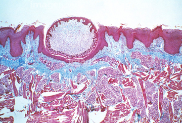
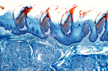
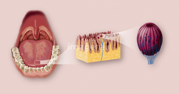
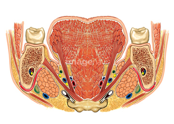
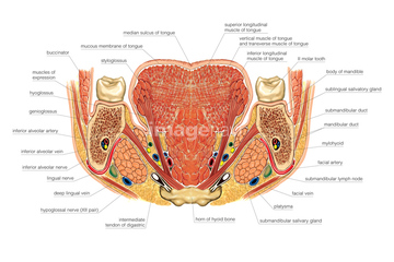
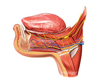
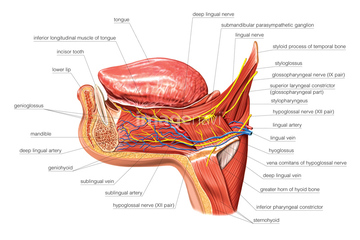
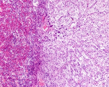
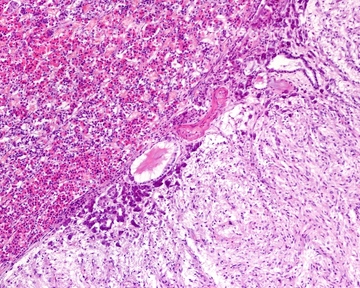
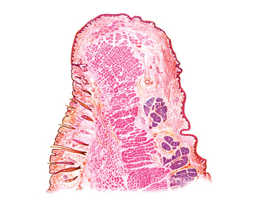
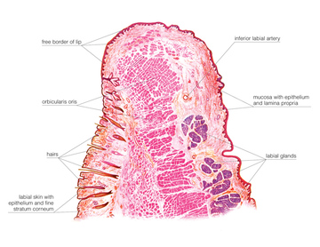
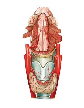
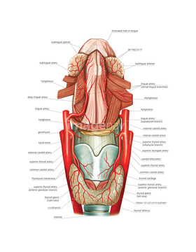
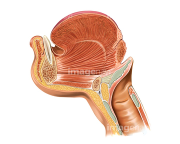
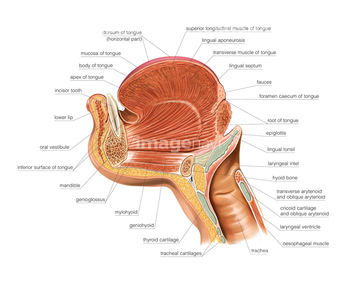
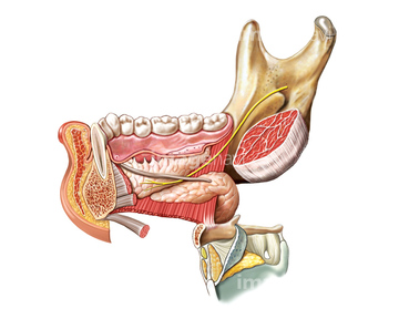
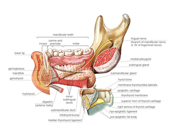
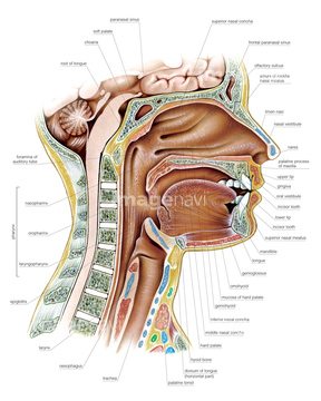
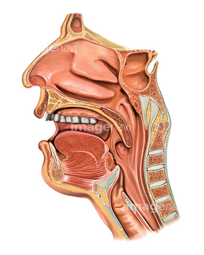
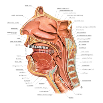
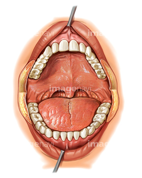
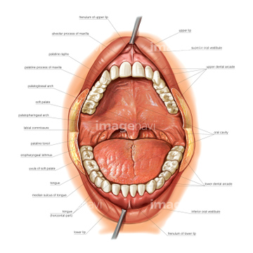
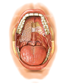
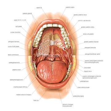
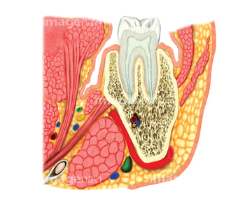
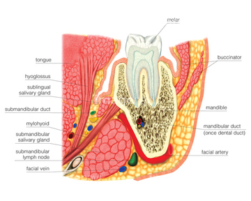
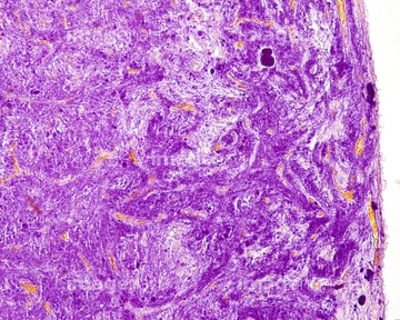
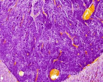
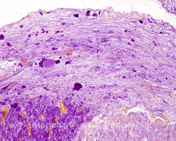
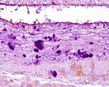
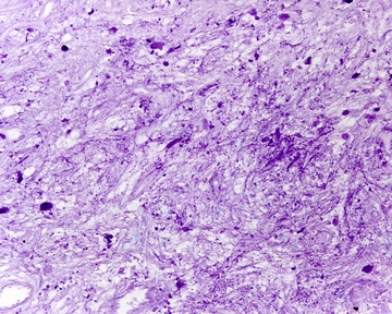
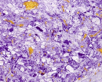
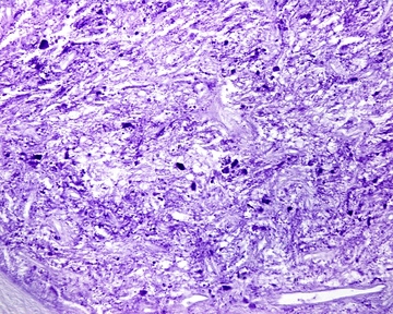
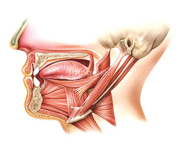
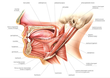
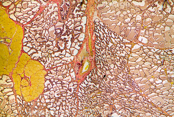
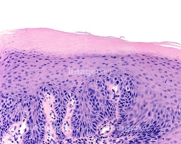
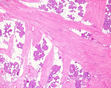
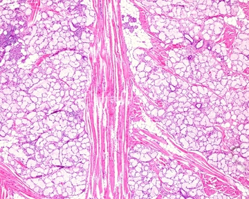
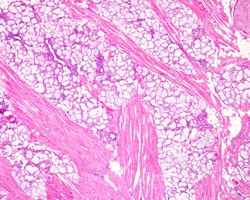
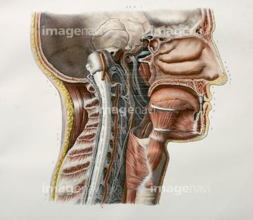
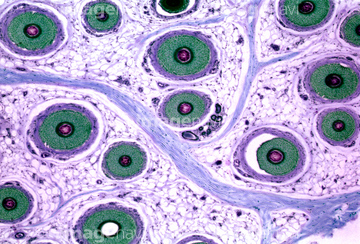
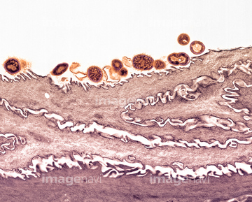
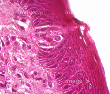
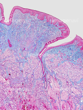
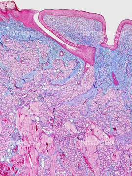
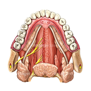
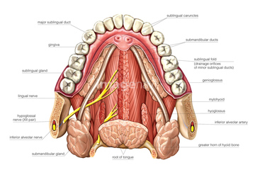
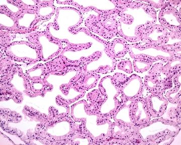
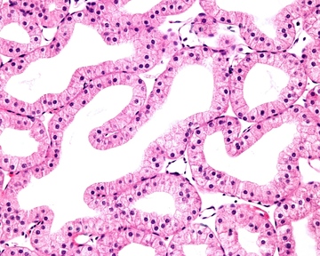
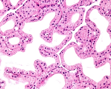
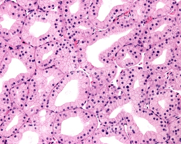
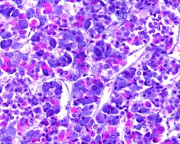
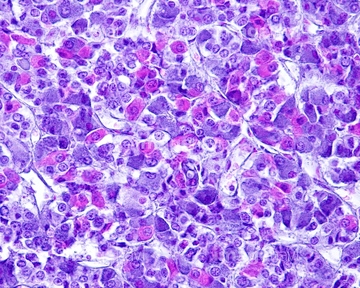
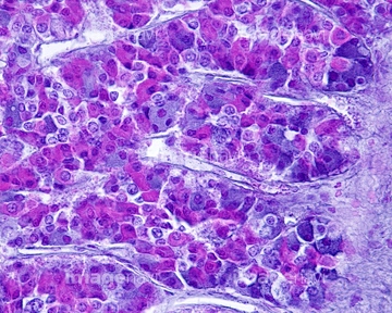
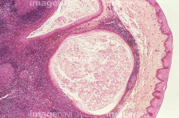
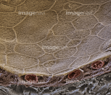
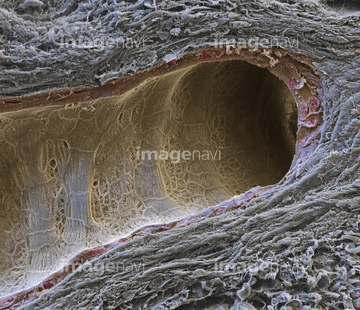
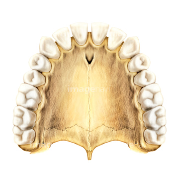
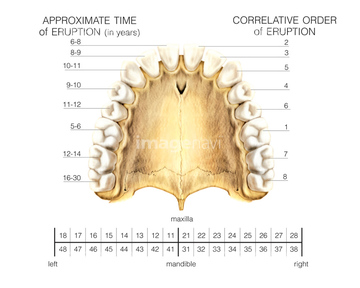
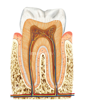
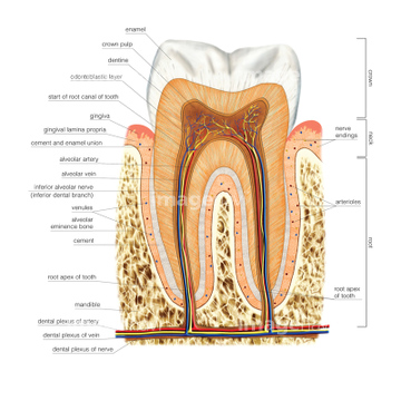
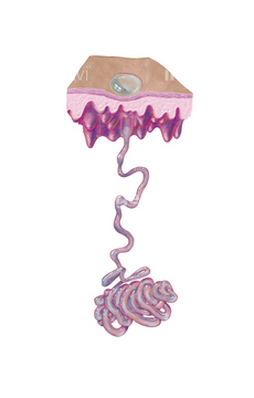
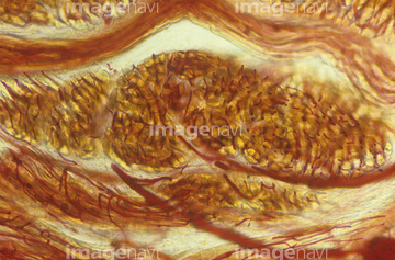
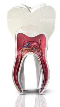
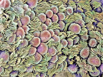
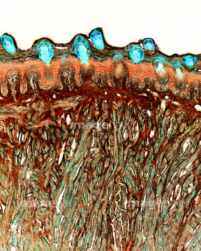
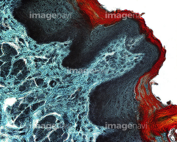
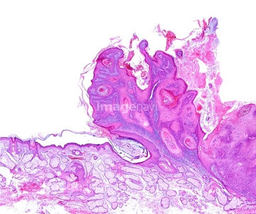
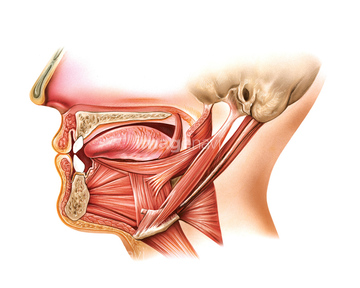
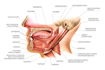
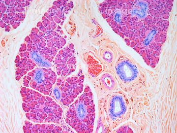
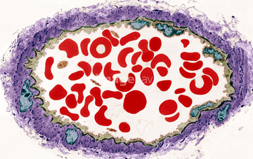
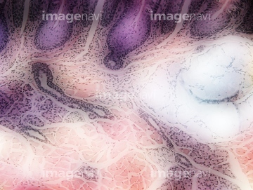
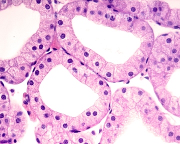
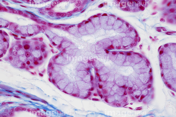
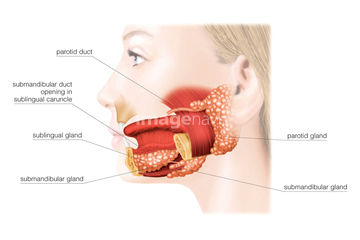
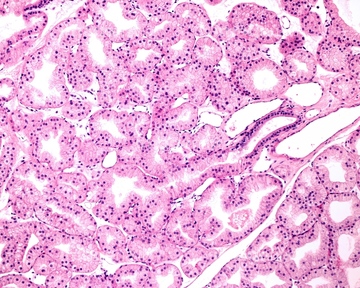
この検索結果には、Digestive System, artwork、Oral cavity and Pharynx, artwork、Oral cavity, artwork、Structure of a molar, artwork、Posterior pituitary gland, light micrograph、Pituitary gland stalk, light micrographなどが含まれています。
17201420
17201421
64050501
64071767
64071768
64071769
64071770
64071771
64071772
64235257
64071775
64071776
64071783
64071784
64206779
64206780
64206781
64235255
64235256
64071773
64071774
64071785
64071786
64071787
64071788
64056743
64235062
64235063
64047103
64047104
64060068

64071753
64071754
64071755
64071756
64071757
64071758
64071759
64071760
64071761
64071762
64071821
64071822
64231753
64070324
64070325
17200010
17200011
64206784
64206785
64206786
64013159
64146222
17201561
64022070
64234649
64049912
64049018
64146190
64146196
64235060
64052161
17201562
64003130

64003131
17206961

17206860
64067718
64070210
64070211
64235181
64235182
64071765
64071766

64022470
64235183

64234840
64234841
64234843
64092797
64092798
64189464
64189466
64189467
64189468
64189469
64189470
64189471
64189472

17201434
64068329
17206960
64060357
64231733
64092746
64176897
64176899
64071805
64071806
64071817
64071818
64071819
64071820
64071825
64071826
64060069
64225232
64225233
64072837
64196719
64059428
17201435
64180242
64180243
17200013
64048781
17201378
64169967
64197460
64071781
64071782
17235967
17201382
64139404
64198946
64235099
64235102
64070284
64070285
64071793
64071794
64189465
| 次ページ |
