HOME > 写真 > 科学・テクノロジー > 科学 > DNA・細胞
10,000件の写真素材が検索されました。
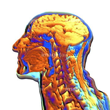
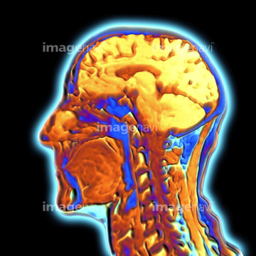
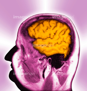
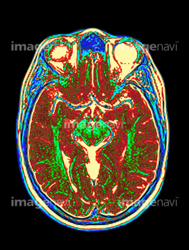
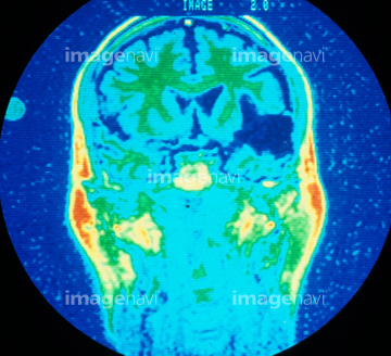
この検索結果には、Coloured MRI scan of the human head、Normal brain, MRI scan、MRI scan of woman's brain (Digital Enhancement)、Normal head and neck, MRI scan、Normal head and brain, MRI scan、Vascular system of the head, MRI scanなどが含まれています。
17206954
17206955
30064857
64064693
64064694
17206956
64021474
64021475
64021476
64021477
64021481
17201401
17201404
64021468
64021469
64021470
64021471
64021472
64072632
64072640
64072641
17206952
64021464
64043086

64043087
64043088
64043089
64043090
64043097
64043098
64043099
64043100
64043101
64043102
64043103
64043104
64043105
64043106
64043107
64043108
64021473
64021478
64021479
64040391
64040392
17218191
17218192
17218197
17218200
17218204

17218210
17201407
17201776
17201768
64014403
64049872
64021454
64043091
64043092
64043093
64043094
64043095
64043096
64021451
64021452
64021453
64021456
64049873
64021447
17201739
64075025
64075026
64075027
64075028
64021444
64021450
64021466
64021467
64066826
64066827
64066833
64066834
64066835
64163477
64043117
64043118
64043119
64043120
64043121
64043125
64043128
64043129
64043130
64263774
64021465
64043131
64063379
64021455
64021443
64021445
17218208
64078939
17223829
17223837
17223840
10941464
64078080
64078081
30342234
17260856
16918270
16918279
16918297
16918323
16918325
16918332
16918335
64078940
64071942
64078941
64021446
64021448
64035457
64035458
64161164
64161165
64161171
64161172
64161174
64161176
64161177
64161178
64161179
64161181
64161182
64161183
64161187
64161194
64161200
64161201
64161204
64172494
64172495
64172496
16918314
16918315
64187413
64042984
64071925
64071926
64071927
64059701
64071706
64071707

| 次ページ |
