HOME > 写真 > 人物 > 構図 > 横向き
10,000件の写真素材が検索されました。
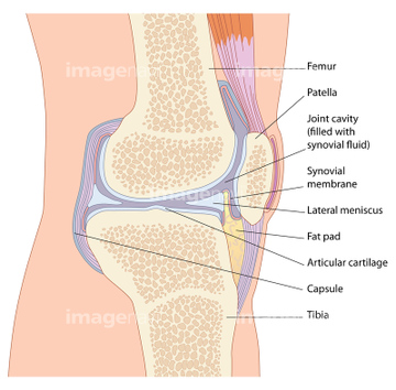
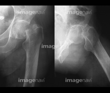
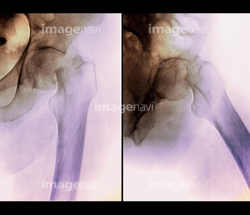
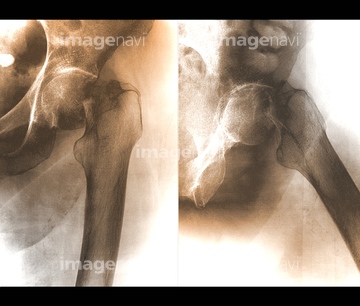
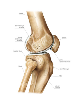
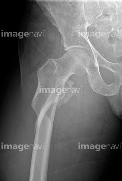
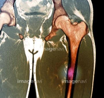
この検索結果には、Broken hip, X-ray、Fractured hip joint, X-ray、Healing broken hip, X-ray、Knee joint anatomy, artwork、Hip stress fracture, MRI scan、Colour CT scan: broken thigh bone in osteoporosisなどが含まれています。
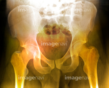
17237155
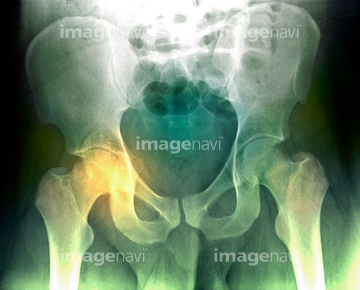
17237144
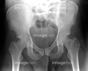
17237145
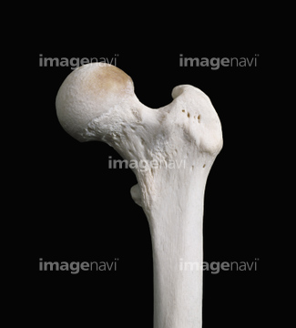
17237158
17237154
17237149
17237156
17237157
17237153
17237147

64225118
64225003
64109925
64109924
64225382
64225007
64225455
64225117
20559313
20559314
20559315
20559316
20559317
20559318
20559319
20559320
20559321
20559322
20555788
64014105
64045499
64045500
64045501
64225369
64225391
64225397

64225370
64224980
64225298
64209196
64209197
64209198
64209199
64209200
64137953
64196406
64049302
64221968
64221969
64221970
64168725
64168726
64168727
64168728
17200502
20573700
64225468
64225469
20512550
20512551
64045834
64071693
64071695
64071696
20573699
64250019
64011419
64012620
64012621
64196405
64100527
64012562
64072124
64012622
64012623
20573694
64168529
64168530
64050519
17200501
64012992
64059505
64064639
64064640
64071951
64071954
20573695
20573696
64219700
64219701
64219702
64219703
64219704
64219705
64219706
64219707
64219708
20512552
20512553
64137954
17249896
17249897
17249898
17249899
20573692
20573697
20573698
64224999
64018453
20512554
20512555
20512556
20548420
17261846

17261847
64012619
64012607
64012608
64012614
64012615
64010066
64056862
64064637
64064638
64064641
64049300
64049831
64049832
64049847
64049874
64040050
64070821
64070822
64071469
64071470
64074821
64057264
64057265
64057271
64057848
64057849
64057850
64234738
64234739
64234740
64234741
20512549
64128017
64159939
64159940
64236964
64261193
64261194
64109932
53122342
53122377
53122649
64013810
64013813
64013814
64013815
64070807
64070808
64070809
64070810
64070811
64070812
64070813
64070814
64070815
64070816
64070817
64070818
20573693
64168531
| 次ページ |
