HOME > 写真 > イラスト・CG > 医療 > 細胞
10,000件の写真素材が検索されました。
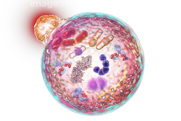
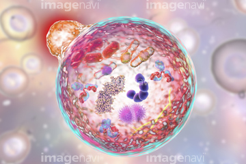
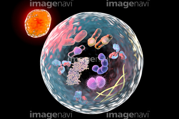
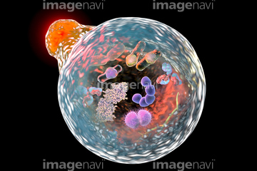
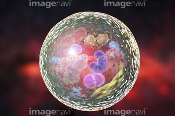
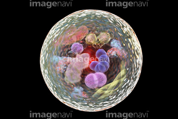
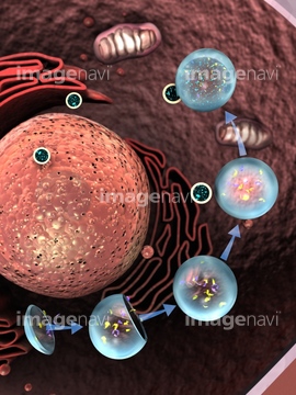
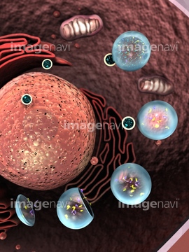
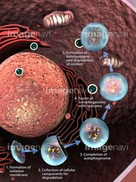
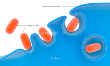
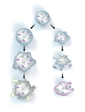
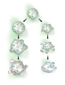
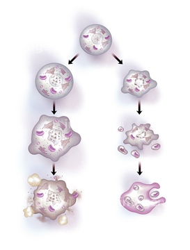
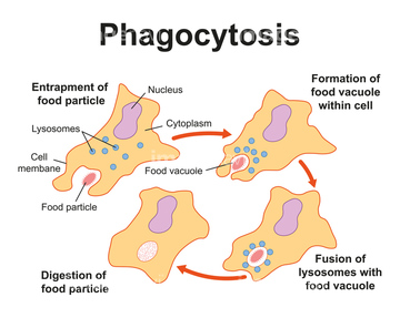
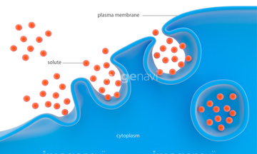
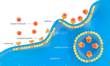
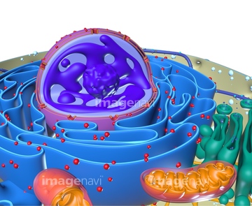
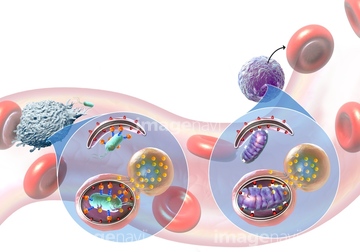
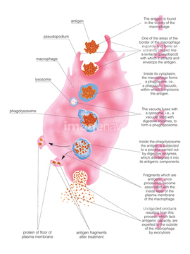
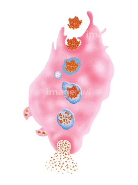
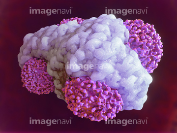
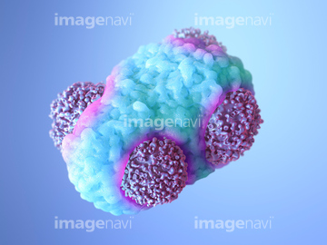
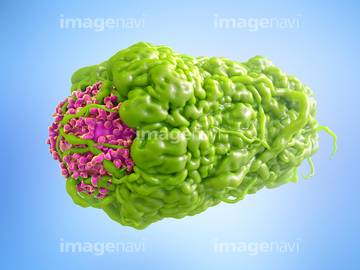
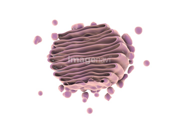
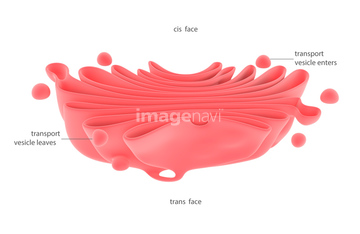
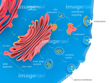
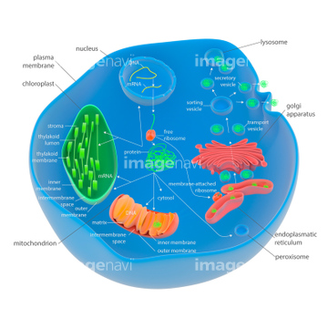
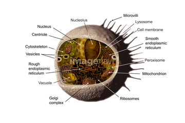
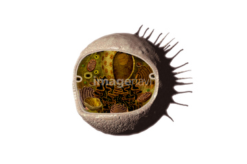
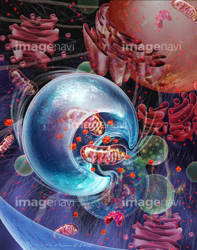
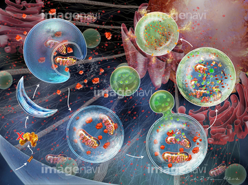
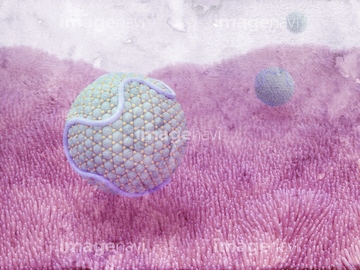
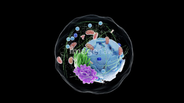
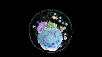
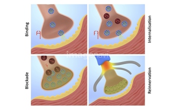
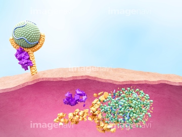
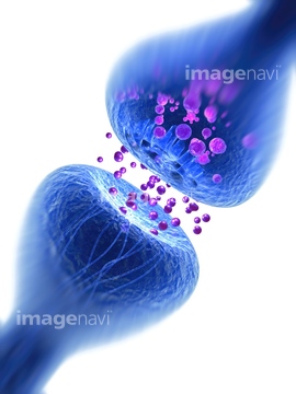
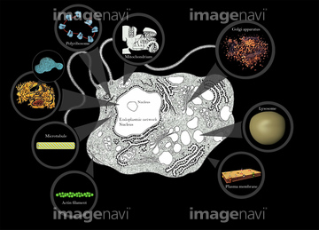
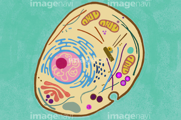
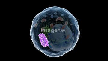
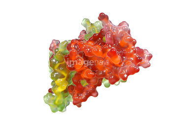
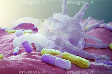
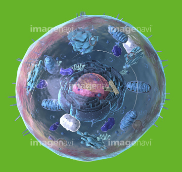
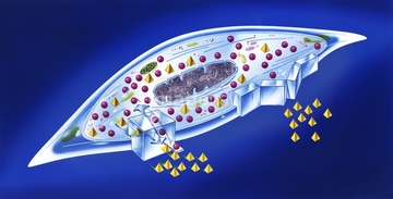
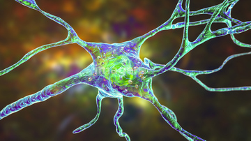
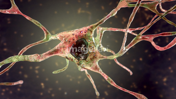
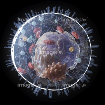
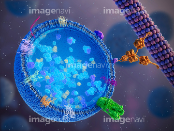
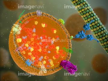
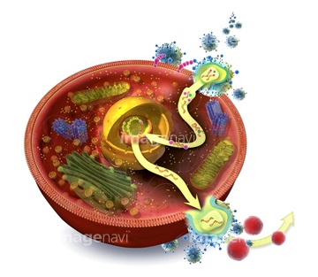
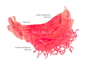
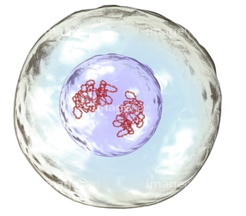
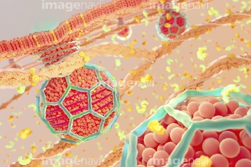
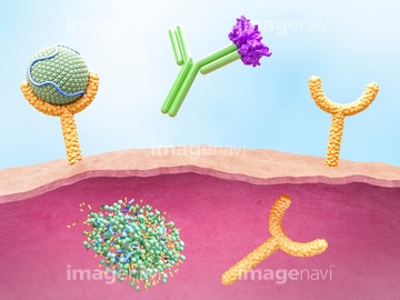
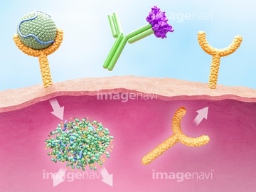
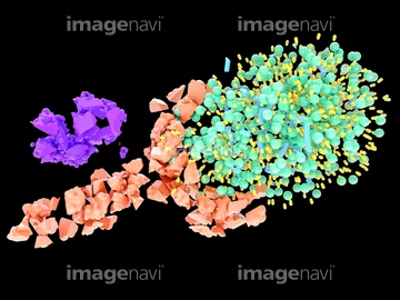
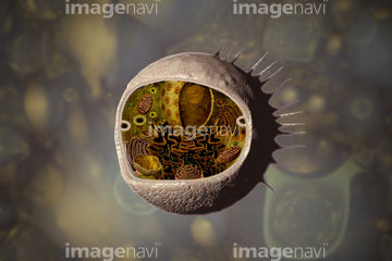
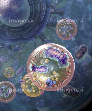
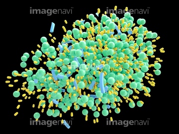
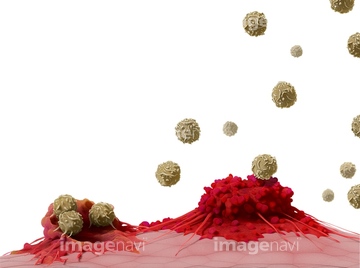
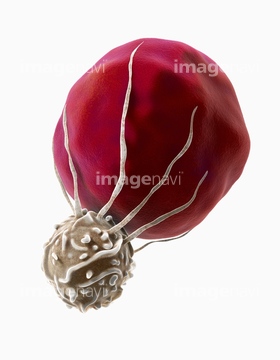
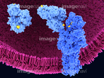
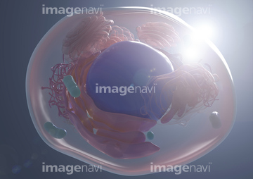
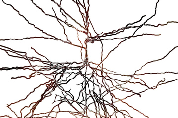
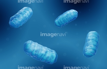
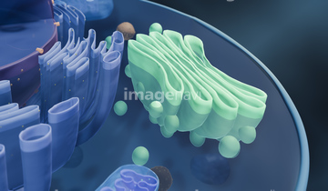
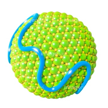
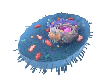
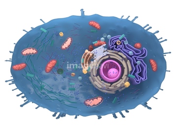
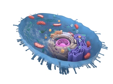
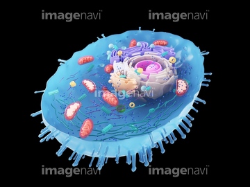
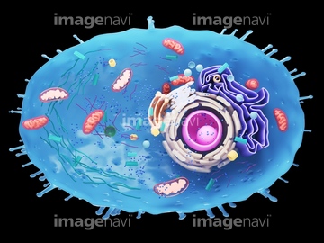
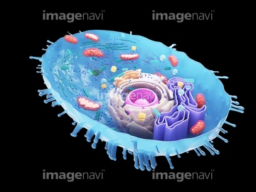
この検索結果には、Macrophage engulfing cancer cell、Monoclonal antibody ipilimumab and receptor, illus…、Human cell, illustration、Lysosome, illustration、Golgi apparatus, illustration、Cellular protein transport, illustrationなどが含まれています。
17257722
17257721
17257715
17260310
17257714
17257716
17257717
17257718
17257719
17257720
64129790
64129791
64148194
64089983
64128498
64128499
64128500

64128501
20571215
64089984
64217190
64217401
64089952
64256978
64063430
20547387
20547545
20547546
20547548

64089989
64131140
20573255
64105844
64129619
64129622
64201656
64201657
64120821
64120822
64097797
64097798
64097799
64097800
20577151
20541225
17270723
64089960
64112599
64118099
64168698
64089953
64137903
64137904
64089650
64089651
64144473
64186161
20577149
20577150
64178029
17264071
17264077
64181149
64264121
64247499
20577153
64245133
64123424
64256977
64194378
20546301
20546302
17288584
20573252
20573253
64089982
64090022
64090023
64144315
64089958
64165068
64137906
64168717
31040757
20571229
20571230
20571231
20571232
64110000
64099415
64085897
64085898
64085899
64085900
64085901
64085902
64085903
64085904
64085906
64085907
64085938
64252110
64047182
64201655
64105843
20573495
64179293
17293923
17293926
17293934
40551584

40551585
64144590
64182428
64182430
64182435
64182437
64179229
17277997
17277998
17294339
17294340
64217189
17298692
20544253
20544281
20544282
20544283
64168694
64160247
64263459
64263460
20573526
64087439
20540498

40561108

40561118

40561119

40561120

40561121
17270536
40545789
64173699
64173700
64097394
64218377
64090044
| 次ページ |
