HOME > 写真 > 医療・福祉 > 医療 > 医薬品
10,000件の写真素材が検索されました。
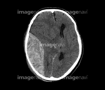
この検索結果には、Carcinoma cell, TEM、Lung cancer cells, SEM、Fibrosarcoma tumour、Extradural haematoma, CT scan、Tumour, artwork、Nanobot detroys cancer in human bodyなどが含まれています。
64054760
64054761
54003423
64211182
17200422
17200423
64013006
64210634
64263204
20559265
64054762
64054763
53157292
53157293
53157294
53157295
53157296
53157297

53157298
53157299
64094074
64150294
64202839
64244160
64244206
30078466
64253396
64122795
64122796
64122797
64122798
64122802
64152580
64043469
64184370
64184371
64201362
64096074
64096084
64203426
64203431
64203451
17217755
17217756
64221246
64246760
64049962
64088501
64261243
64261244
17212310
17212311
64052113
64097123
17274515
17274516
17274518
64207259
64207269
17201296
17201297
64055690
64063397
64219707
64219708
64158222
64158234
64158253
64158267
64195828
64195841
64195860
64221074
64155659
64155691
64221196
64221197
64221198
20564518
53156654
53156655
53156849
53156850
64152561
64106466
64164114
64164260
64205881
64207250
64212560
64222778
64256046
64256047
64256048
64256049
64256050
64256051
64256052
64256053
64257159
64257160
64257232
64263085
64263133
64263203
64244169
64244172
64244185
64250953
64250954
64250955
64250956
64250957
64250958
64250959
64250960
64250961
64250962
64250963
64250964
64250965
64250966
64250967
64202837
64064331
20582151
20582152
20582153
20582154
64050774
64246761
64246762
64246763
17236831
64225107
53157373
53157374
53157375
53157376
53157377
53157378
53157379
53157380
17236827
17290930
17234586
64176062
64176074
64176076
64176077
| 次ページ |
