HOME > 写真 > 乗り物・交通 > 海路・水路 > 運河
10,000件の写真素材が検索されました。
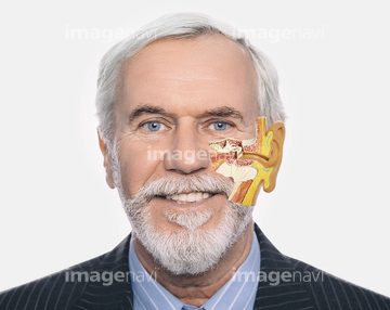
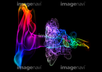
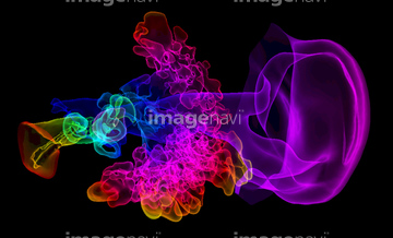
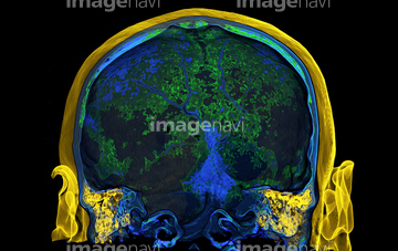
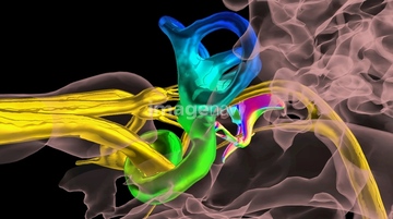
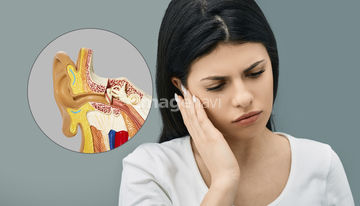
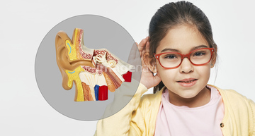
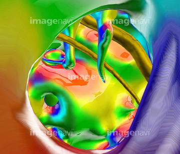
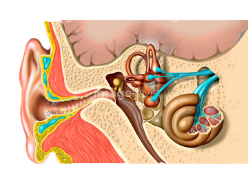
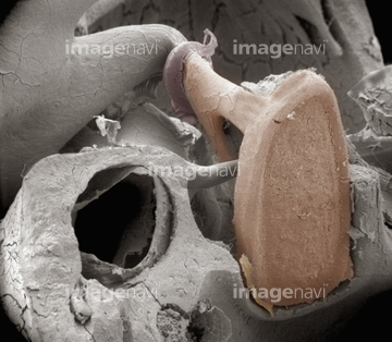
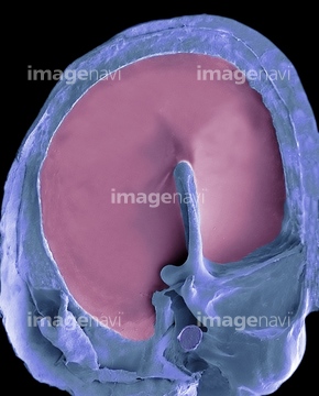
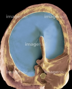
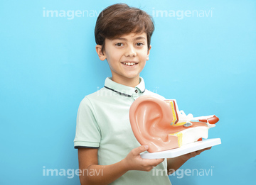
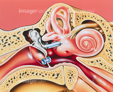
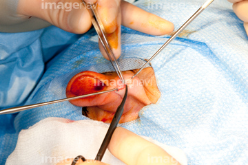
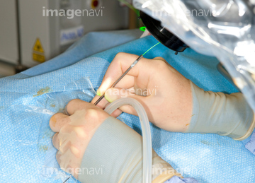
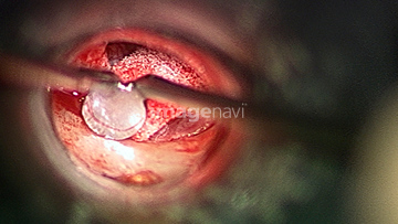
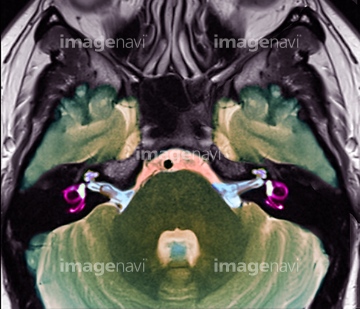
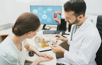
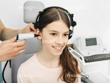
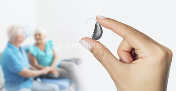
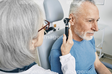
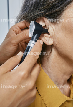
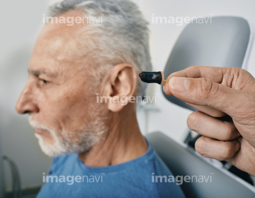
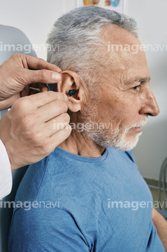
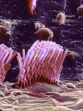
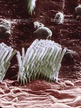
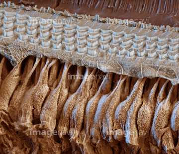
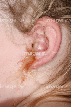
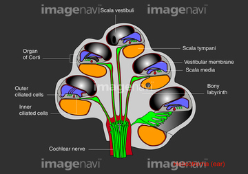
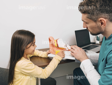
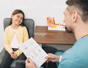
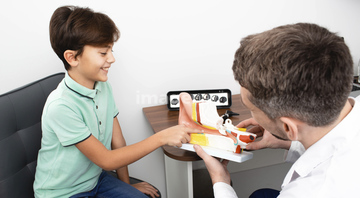
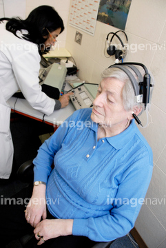
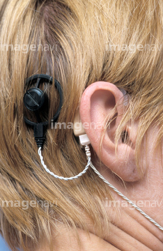
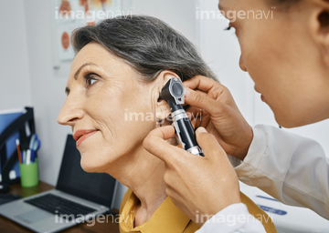
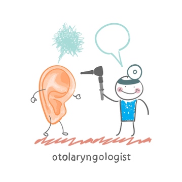
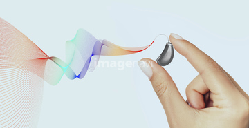
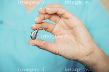
この検索結果には、Ear examination、Axial section of the brain and inner ear, MRI scan、Audiology consultation、Hearing test、Hearing aid、Inner ear anatomy modelなどが含まれています。
20563023
64202541
64202542
64250856
64250857
64080566
64225063
64265185
64265186
64265187
64265188
64265189
64265190
20566235
20563024
64265183
64265184
64220308
64225195
64052109
64225197
64244789
64244828
20560042
20566114
20566115
20566116

20547328

20549080

20549081

20549082
64043308
64043311
64043314
64043315
20546392

20546393

20546394

20546395
20546396

20546397

20546398

20546399

20546400
20546413
20562631
20549164

20560346

17246278
17246279
20546416

20546417
20547599

20549457
20560202
20560203
20560285
20560339
64112209
64043309
64043312
64043313
64049984
64205141
64194305
64194351

20566117

20566118
20566128
20566129

20549083
20549084
20566135
20566130
20566131

20566132
64193341
64019686
20549115

17201317

17201318
20559943

17292052
64167045
64167050
64167051
64167052
64199145
64262939
64262940
20542286
20542287
20542289
20542290
20542291
20542292
20542296

20503236
20503237
20503238
64172333

17203473

17203582
20559976
20560007
20560028
64187455
64187456
64187457
64187458
64187459
64187460

64188008
64188009

64188010
64188011
64008492
64011566
64220309
20566119
20566133
20547330


29093055
20559942
20560200
20560204
20560207
20560209
20560210
20560214
20560215
20560217
20560218
20560284
64123151
64123153

20559961

20559977

20560283
64088778
64192540
64192541
64192542
64198228
64198229
64205102
64205103
64205104
64205115
64205116
64205117
64205118
64205119
64205120
64205121
64205122
64205123
64205124
64205125
64205126
64205127
64205128
64205129
64205130
64205131
64205132

20548682

20549202
20546410

20546411
20546412
17201417
64205071
64205072
64205073
64205074
64205075
64205076
64205077
64205078
64205079
64205080
64205081
64205082
64205083
64205084
| 次ページ |
