HOME > ژتگ^ > ژY‹ئپEٹآ‹«–â‘è > ƒTپ[ƒrƒX‹ئ > ˆم—أپE•ںژƒ‹ئ
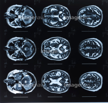
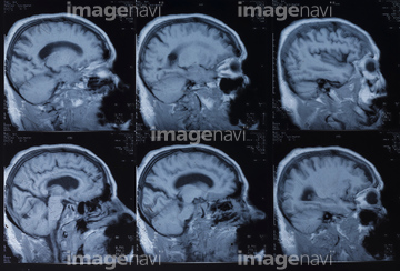
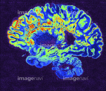
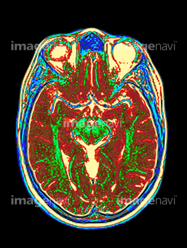
10,000Œڈ‚جژتگ^‘fچق‚ھŒںچُ‚³‚ê‚ـ‚µ‚½پB
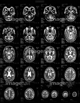
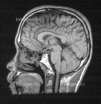
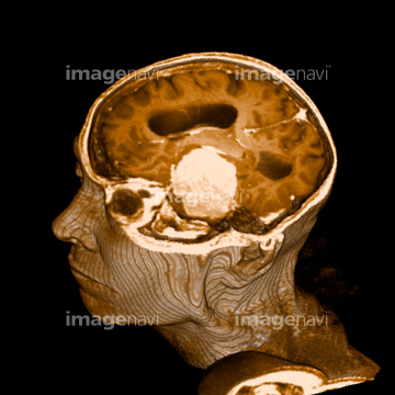
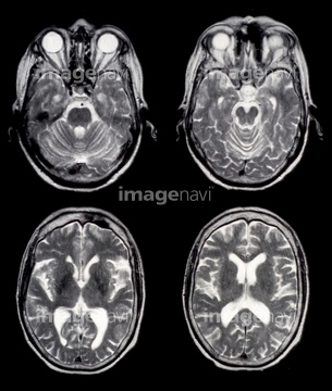
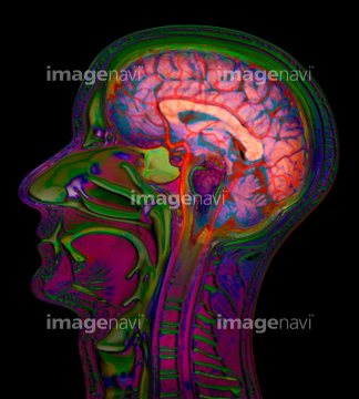
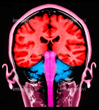
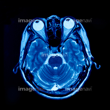
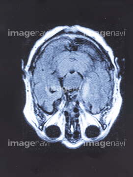
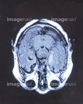
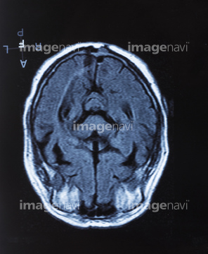
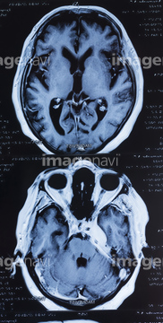
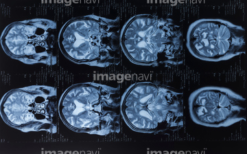
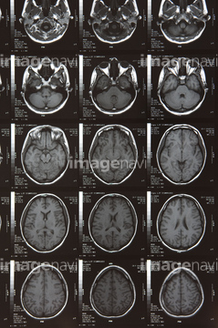
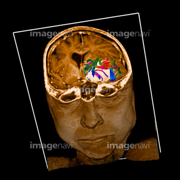
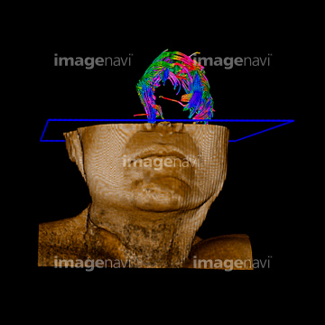
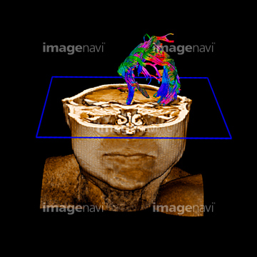
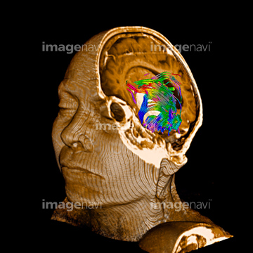
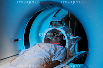
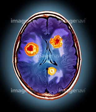
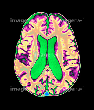
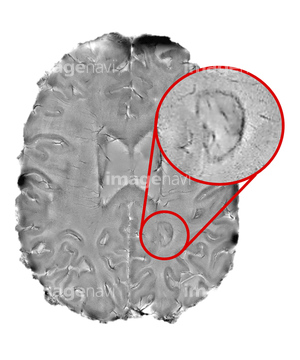
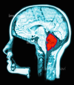
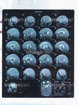
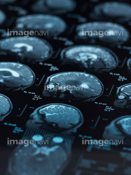
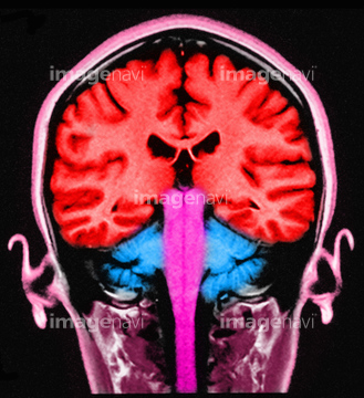
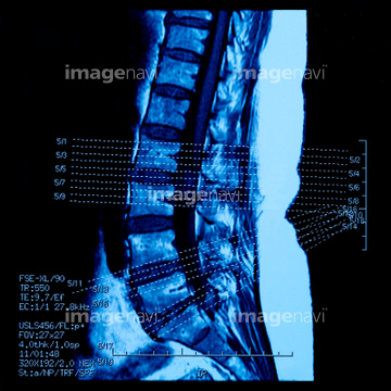
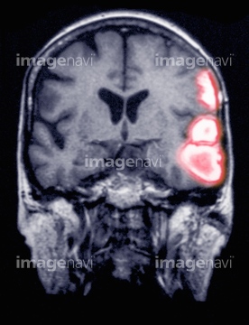
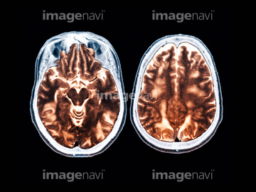
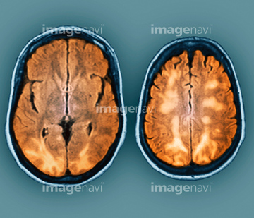
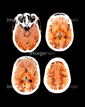
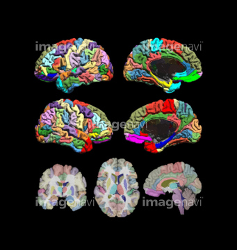
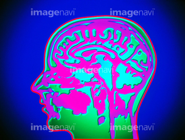
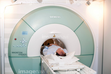
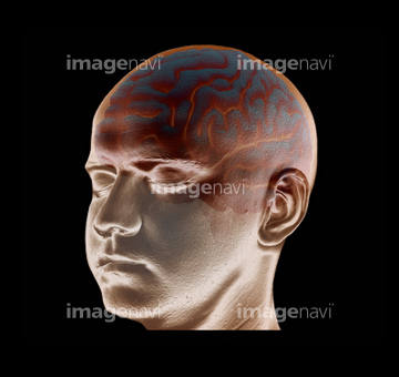
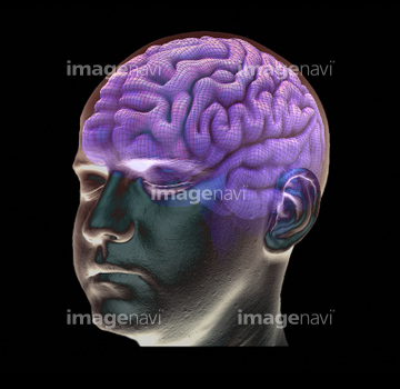
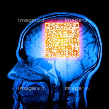
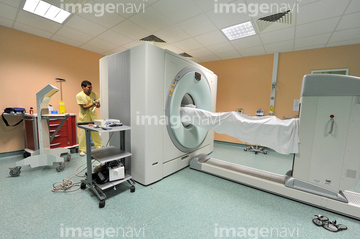
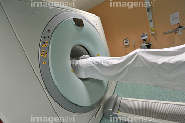
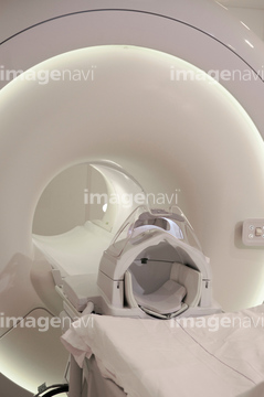
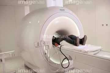
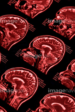
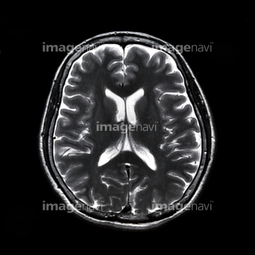
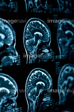
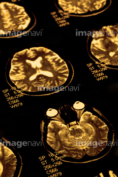
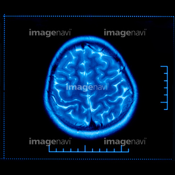
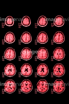
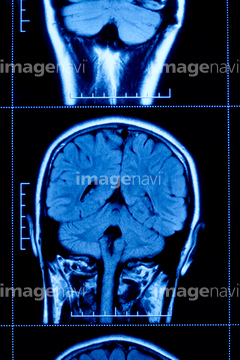
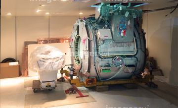
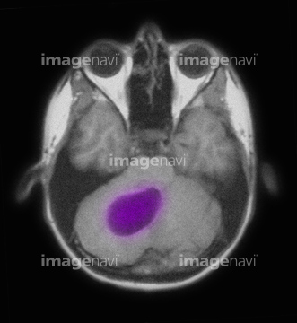
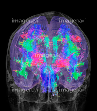
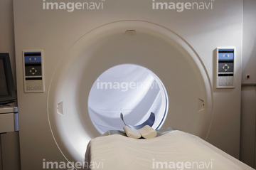
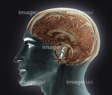
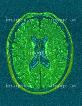
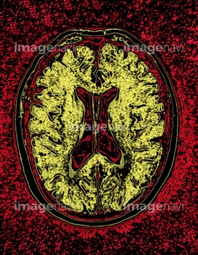
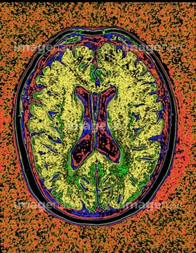
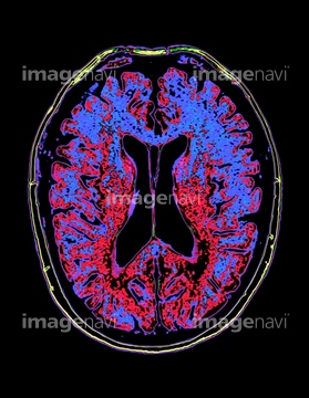
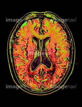
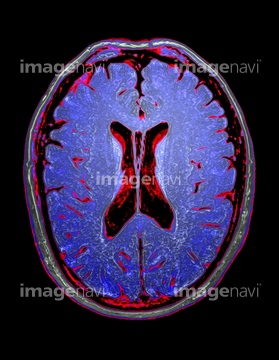
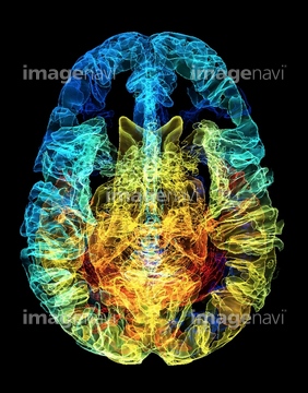
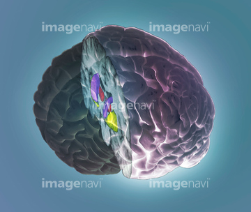
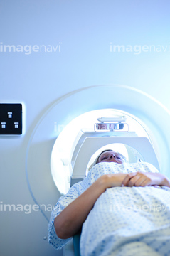
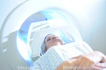
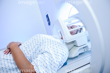
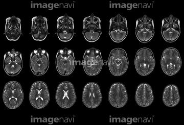
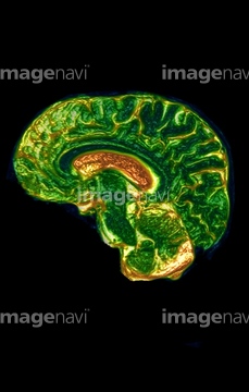
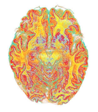
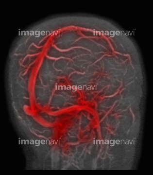
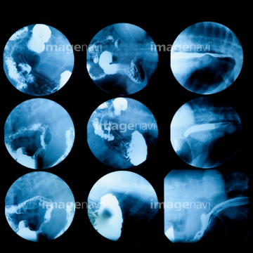
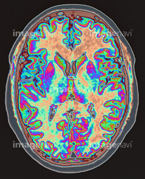
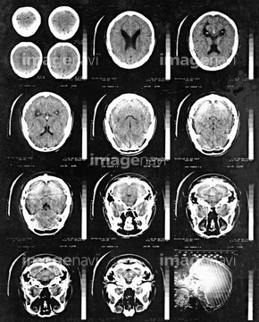
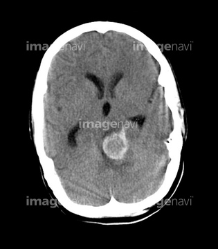
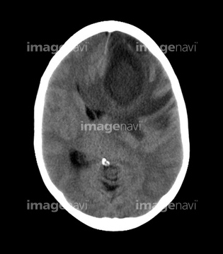
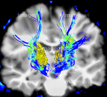
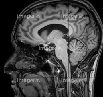
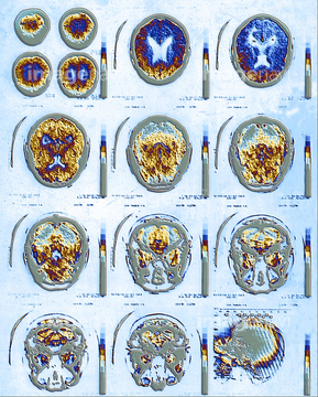
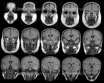
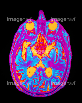
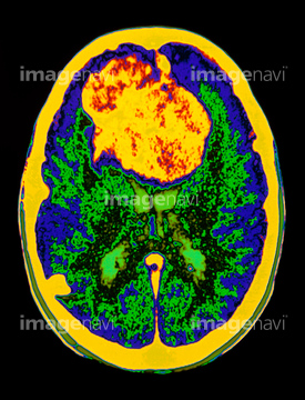
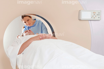
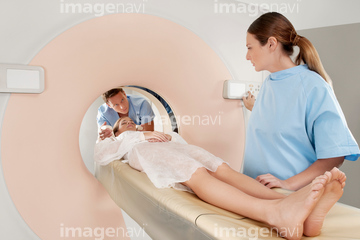
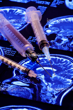
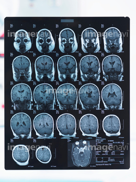
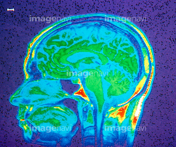
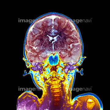
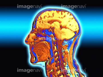
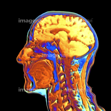
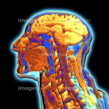
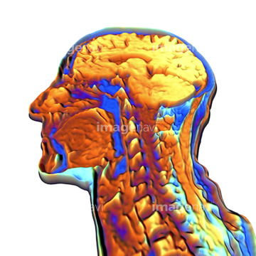
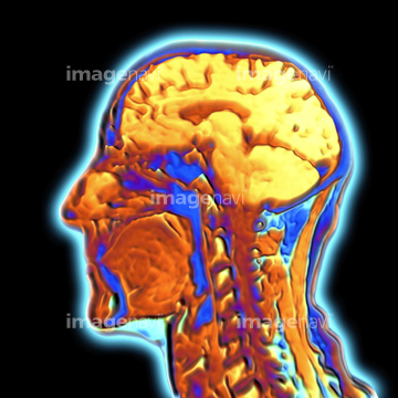
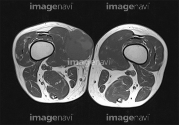
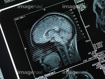
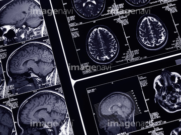
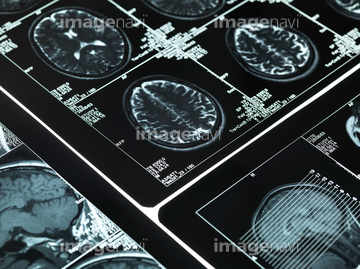
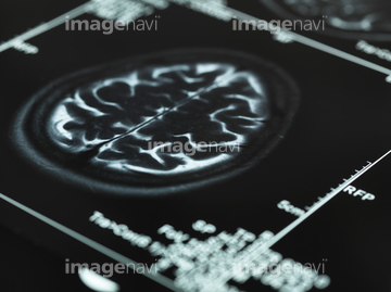
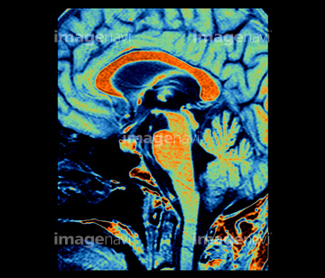
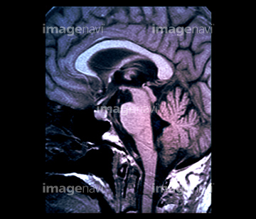
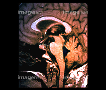
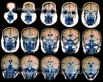
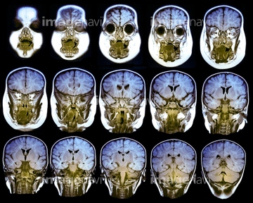
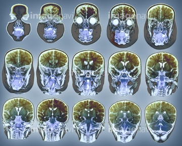
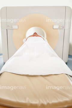
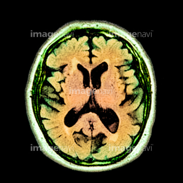
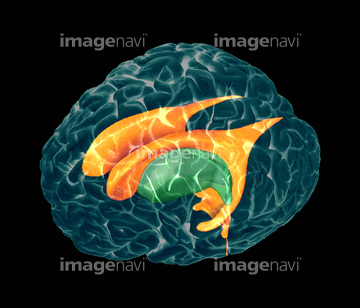
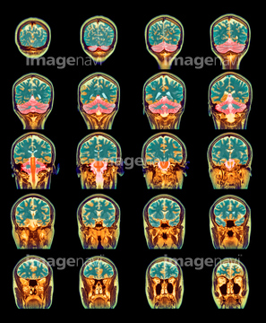
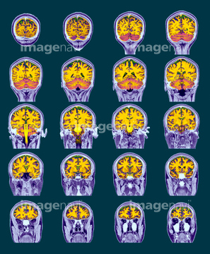
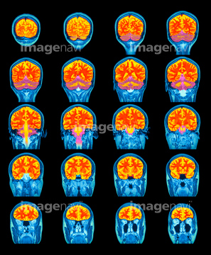
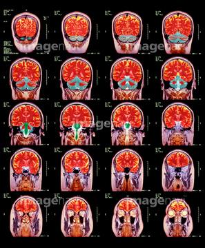
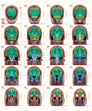
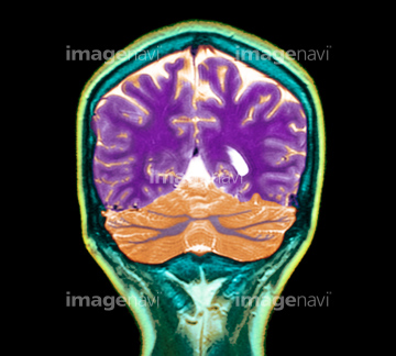
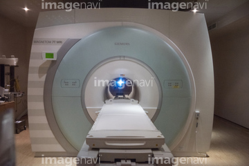
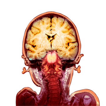
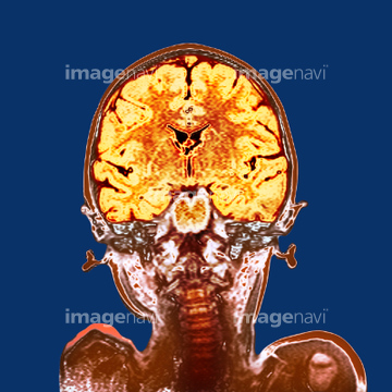
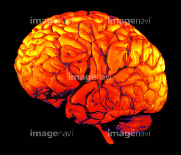
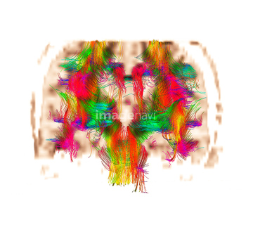
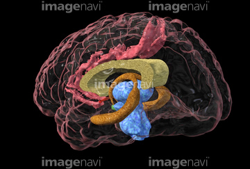
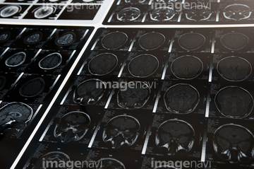
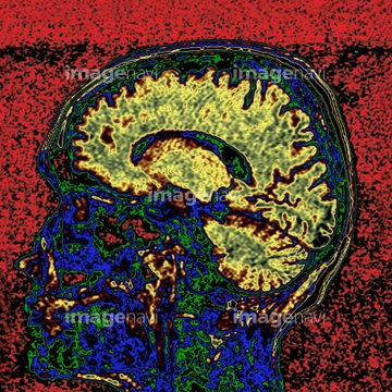
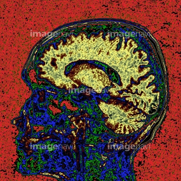
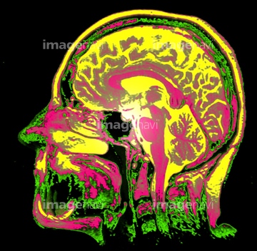
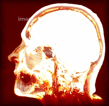
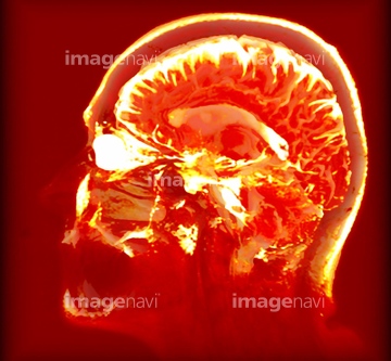
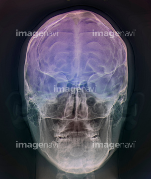
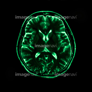
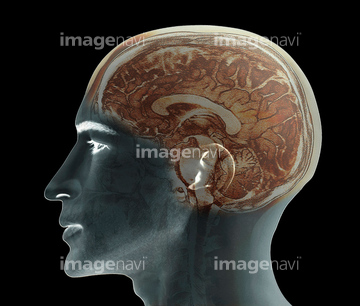
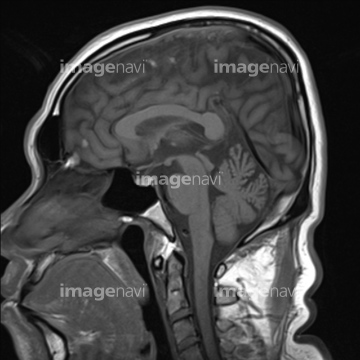
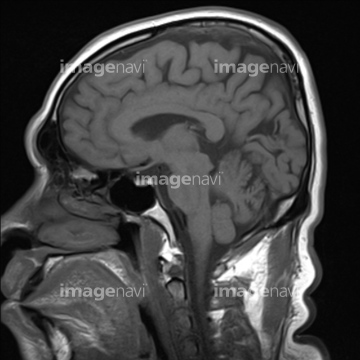
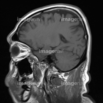
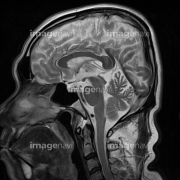
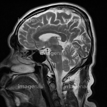
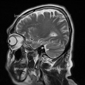
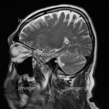
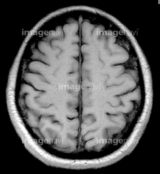
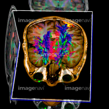
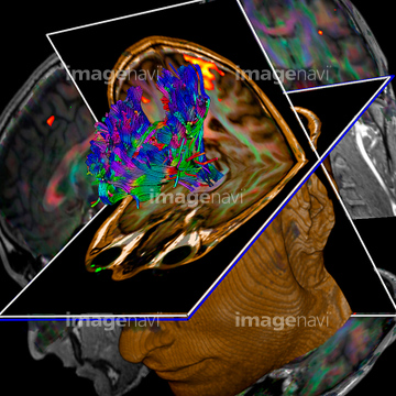
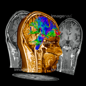
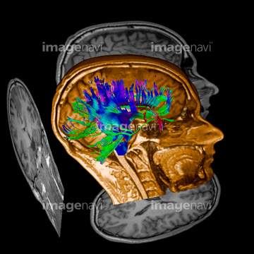
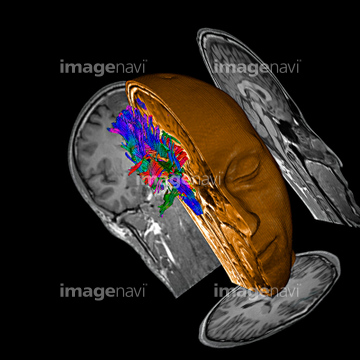
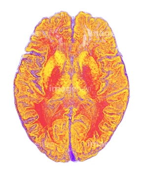
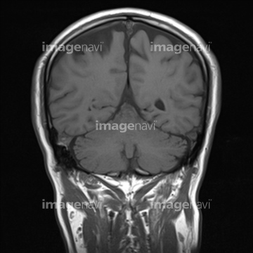
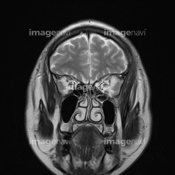
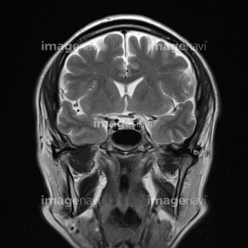
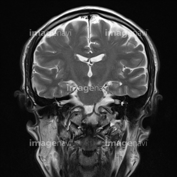
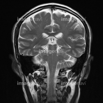
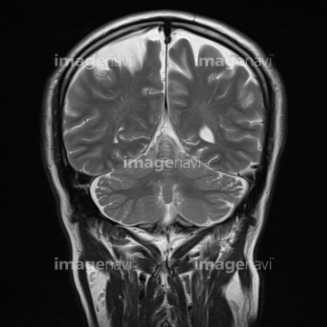
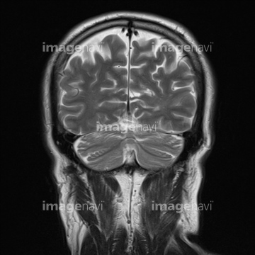
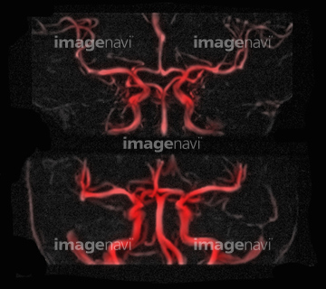
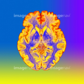
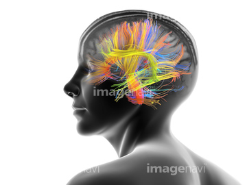
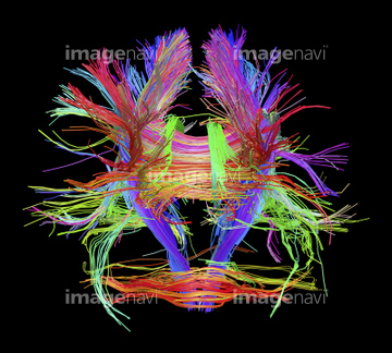
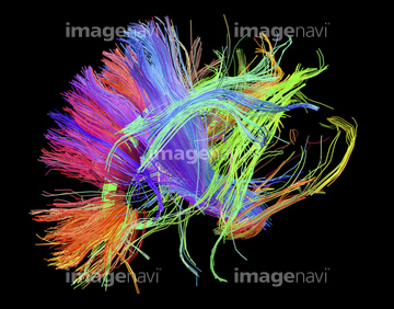
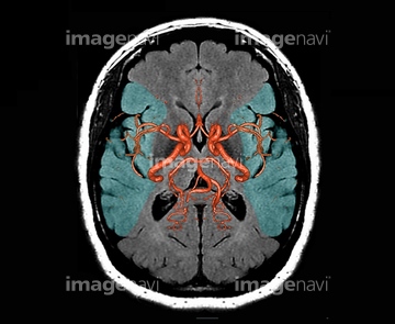
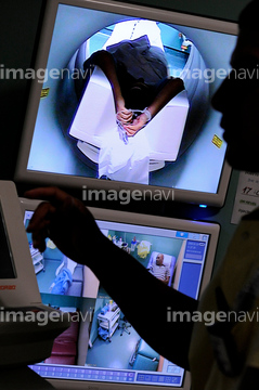
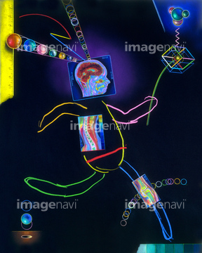
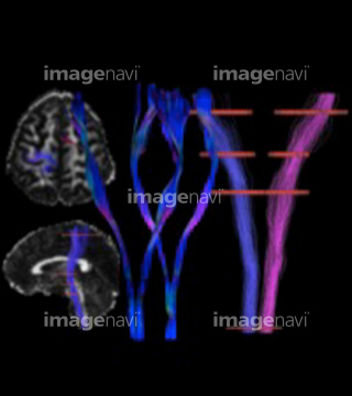
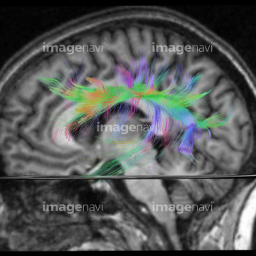
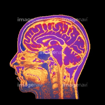
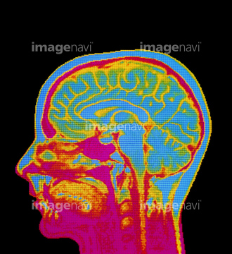
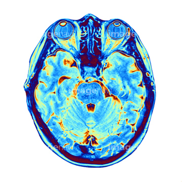
‚±‚جŒںچُŒ‹‰ت‚ة‚حپABrain MRI scan with Alzheimer's QR codeپAMRI scanپAMRI scannerپAMRI scan of human brain, close-upپAMRI scan of brain, close-upپAMRI scan of human head, close-up‚ب‚ا‚ھٹـ‚ـ‚ê‚ؤ‚¢‚ـ‚·پB
64043131
16918270
17280779
17280780
17280781
17280782
17280783
17280784
17280785
64217040
10941464
64043125
64043128
64043129
64043130
64075025
64075026
64075027
64075028
64051975
64049886

20500112
30304357
30304358
20993281
16918273
64188513
64188514
64071942
17201404
64071925
64071926
64071927
64056052
64071706
64071707
64051968
64051969
64052093
64052094
16918314
16918315
16918323
16918325
16918332
16918335
16918297
20566911
64049873
30072385
64064694
64188454
64188455
64188456
64188457
64188458
64188459
64164044
64075285
64062200
64062201

64062202
64062203
64246012
64188464
64149562
66000267
30342233
30342234
64046178
64046179
30342237
64219589
64060094
64060095
66000180
66000580
16918316
30095546
17201401
64014403
17218191
17218192
17218197
17218200
17218204

17218210
21565718
17235936
17235937
17235938
17235939
64245758
64245759
64245760
64220127
64220128
64220129
66000560
64042984
64021454
64021468
64021469
64021470
64021471
64021472
64021473
64110133
64040391
64040392
64021456
64071941
64021447
10941443
64049872
64187413
16918279
16918307
64064693
20573165
20573166
20573167
20573168
20573169
20573170
20573171
30383186
64043117
64043118
64043119
64043120
64043121
64153283
20582138
20582139
20582140
20582141
20582142
20582143
20582144
20582145
20582146
20582147
20582148
64159896
64066833
64066834
64066835
64198138
64198139
64051967
64052088
21508369
64130747
64163476
17218208
64264374
| ژںƒyپ[ƒW |
