HOME > 写真 > 科学・テクノロジー > テクノロジー > バイオテクノロジー
10,000件の写真素材が検索されました。
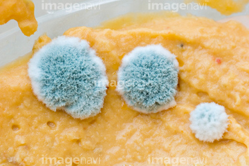
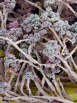
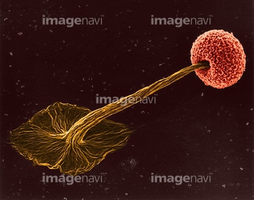
この検索結果には、アオカビ、Microbes growing on agar plate、Plastic surface with bacteria and fungus, SEM、Microbial colonies on petri dish、Microbes growing in Petri dish、Penicillium and bacteria in a petri dishなどが含まれています。
20543469
20543470
20543471
20543473
20544096
20544097
20544100
64003465
64003655
64224853
17275891
17275893
17275906
17275907
17275910
17275911

70014586
64055984

64003661
64003668
64155225
64155227
64003587
64003588
64003589
64003590
64003591
64003652
20543630
20543637
64225097
17278967
17278968
17278969
17278970
17278971
17278972
17279021
17279022
17279023
17279024

17279025
17279026
17279027
17279028
17279097
17279098
17279099
17279100
17279101
17279102
17279124
17279125
20501178
20501179
20501180

20501181
20501182
17283813
17283834
64178273
64178280
64178281
64003653
64003659
64003660
64003667
20545728
20545756
64003609
64003658

20545725
20545726
20545758
64153144
64153158
64128514
64128515
17260791
64003666
64011459
64011460
64106050
64220255
64103031
20569965
20569966
20569967
20569968
20569969
20569970
20569971
20569972
20569973
20569974
20569975
20569976
20569964

64164077
64164078
64164079
20531015
20531016
20531017
64003669
64256625

64256636
64256637
40834040
40834041
40834042
40834043
40834044
40834045
40834046
40834047
64003531
64260981
64260982
64260983
64260984
64098518
64098519
64098520
64003651
64003643
64003646
64003648
64003656
64003664
64003490
64135198
64135199
64135200
64123403
64225084
64055818
17260892
17260893
17261111
17261112
64114581
64107089
64107090
64164075
64164076
64164080
64164081
64164082
64164083

64164084
64164085
64090397
64098511
64098512
64098515
20543533
| 次ページ |
