HOME > 写真 > イラスト・CG > 美術 > アールヌーボー・アールデコ
10,000件の写真素材が検索されました。
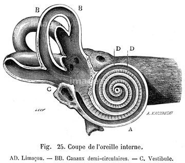
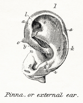
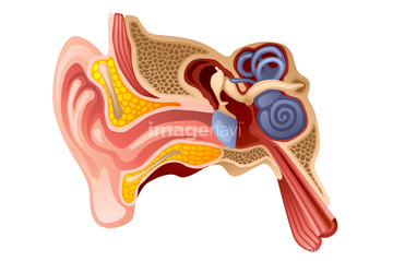
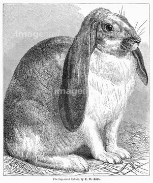
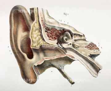
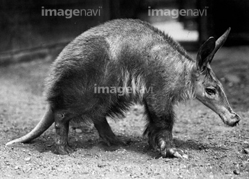
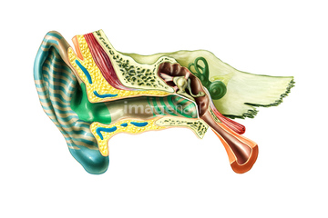
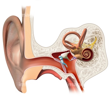
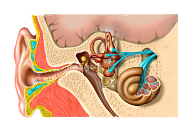
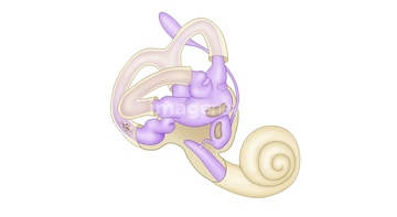
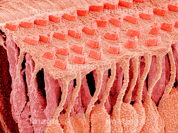
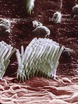
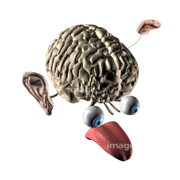
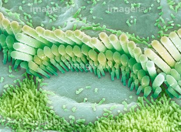
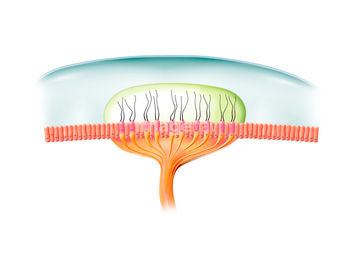
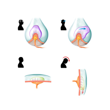
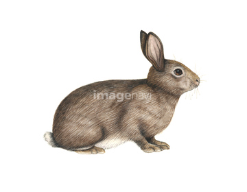
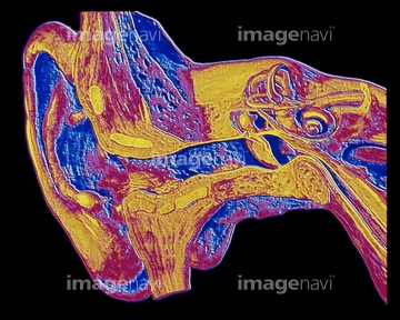
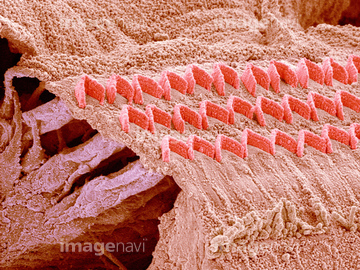
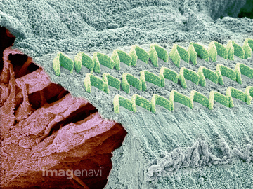
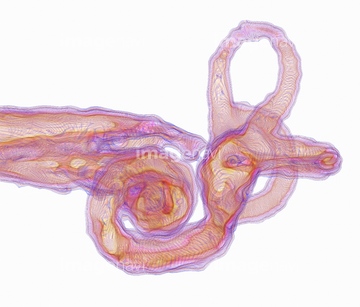
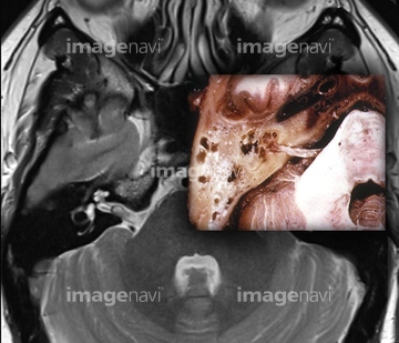
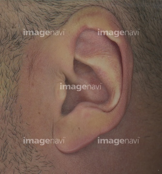
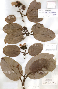
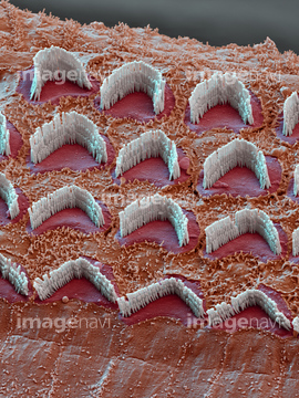
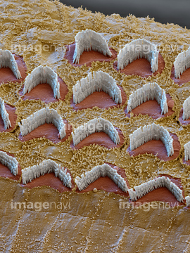
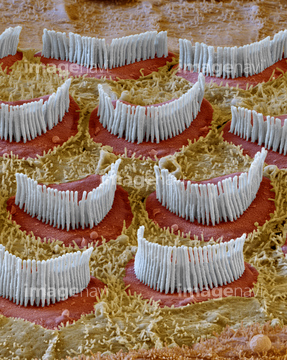
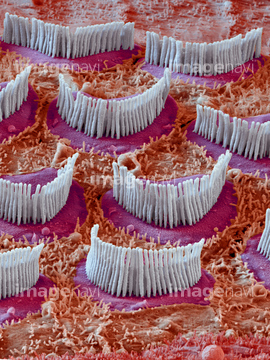
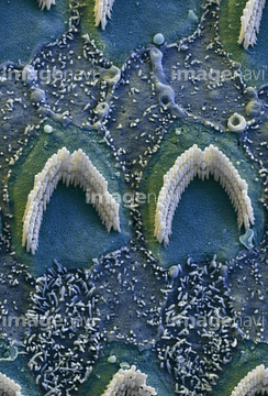
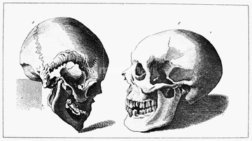
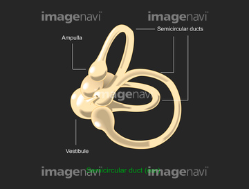
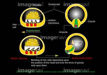
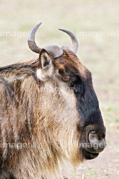
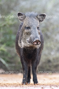
この検索結果には、Inner ear hairs, SEM、Head and neck、Blesbok ram、Ampullary cupula, artwork、Inner ear hair cells, SEM、Sound waves human ear, illustrationなどが含まれています。
51975788
51975783
51970692
51975784
51985453
51975785
51975786
51975787
51975789
51975790
51934469
51934466
51934468
64224220
16929677
52104392
64060310
64044882
51934467
64100259
64100260
64021485
64223638
64052158
52102171
64064269
17207857
17207858
17207859
17208224
64163513
64049984
64039926
17207090

51975792
51912516
51913731
52102785
64200400
64200402
64200408
64200412
64225195
52104372
64009067
64009071
64009072
64009074
64009076
51461299
64059800
64062169
64039927
64039928

64238645
64009056
64074757
17225336
17225340
64113536
64052109
64179285
64050571
64009170
64009171
64225197
64075654
64044818
64220309
64168010
64168011
64167045
64167050
64167051
64167052
64060081
51912517
51914613
52104437
51983829
51426396
64076865
64057359
64057360
64057521
64057522

64008675
64008677
64009051
64009063
64009064
64009065
64074758
64062170
64062171
64043696
64082470
64082471
17206188
64078913
64088778
64009872
17207097
64225070
64035023
64179286
10990982
64076909
64059171
64084946
64153321
64168012
64192565
64199772
64054485
64059060
64051760
64073464
64073465
10983331
10914325
30407945
64205076
64205077
64205078
64205079
64205088
64205089
64205090
64205091
64205100
64205101
10920719
64199145
51490934
64044120
51994173
51975791
51972800
51975798
64190510
64048783
64048790
64043460
64035730
64037391
64185724
64185735
64227425
64219270
64088572
64239415
17201417

64220308
64225069
64064270
10919281
53124199
64076758
52101863
64043486
64049439
64049441
64073471
51952513
51952514
64060993
52102246
64052518
64076809
64076810
64076811
64076813
| 次ページ |
