HOME > 写真 > 科学・テクノロジー > 科学 > DNA・細胞
10,000件の写真素材が検索されました。
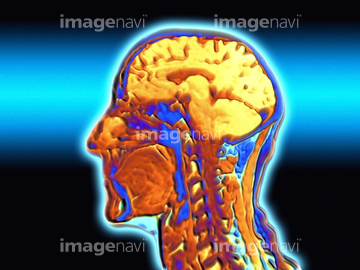
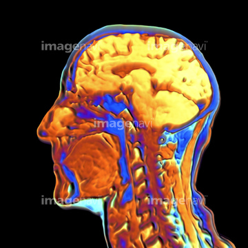
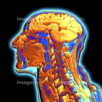
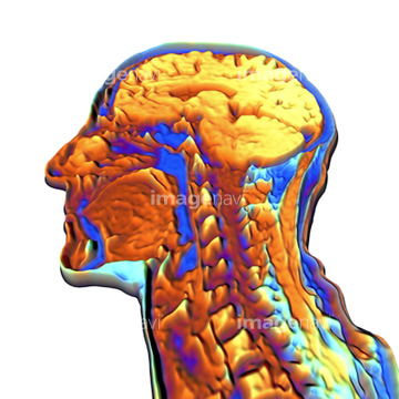
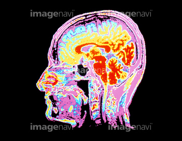
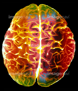
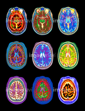
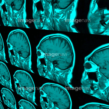
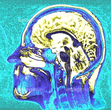
この検索結果には、Coloured MRI scan of the human head (side view)、Healthy brain, MRI scans、MRI scans of the human brain、Coloured MRI scan of the human head、Coloured MRI scan of the human hea、MRI Scan of human head, close-upなどが含まれています。
53156553
53156550
53156551
53156552
53156554
53156555
53156548
53156549
53156556
53156557
53156558
53156559
53156560
53156561
53156562

53156563
64066826
64066827
64066833
64066834
64066835
64051975
21508369
64042984
64043125
64043128
64043129
64043130
64043131
64071925
64071926
64071927
10941464
30342237
64219589
64049886
17201401
17201404
17260856
17218191
17218192
17218197
17218200
17218204

17218210
16918270
16918314
16918315
16918316
16918323
16918325
16918332
16918335
64110133
64161164
64161165
64161171
64161172
64161174
64161176
64161177
64161178
64161182
64161187
64161200
64161201
64161204
64172494
64172495
64172496
64064693
64064694
64049872
64049873
64040391
64040392
64014403
64071447
64071706
64071707
64071942
64075025
64075026
64075027
64075028
64021447
64021451
64021452
64021453
64021454
64021456
64021468
64021469
64021470
64021471
64021472
64021473
64021475
64021477
64021478
64021479

21555491
64114171
17200425
64071938
64071940
64246618
64245956
64046178
64046179
64046182
17218208
64059701
64010056
64021481
64021443
64021444
64021445
64021446
64021448
64021449
64021450
64021455
30064857
64051974
16918297
20562166
64161158
64161159
64161160

64161163
64161179
64161181
64161183
64161194
64161202
64187413
64161161
64161162
| 次ページ |
