HOME > 写真 > 科学・テクノロジー > 科学
10,000件の写真素材が検索されました。
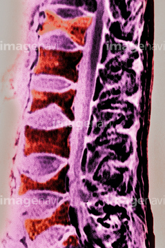
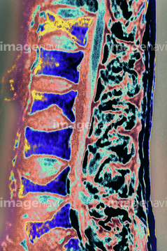
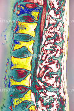
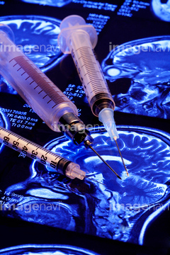
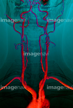
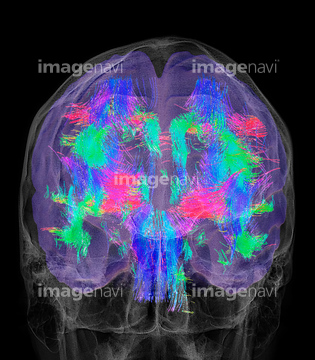
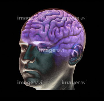
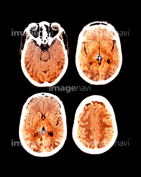
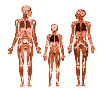
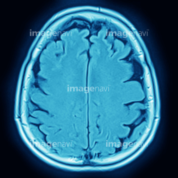
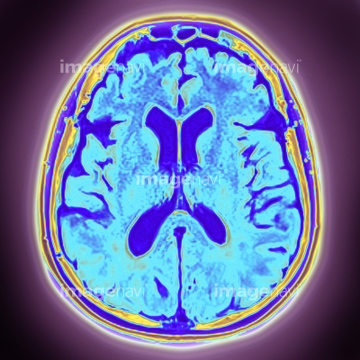
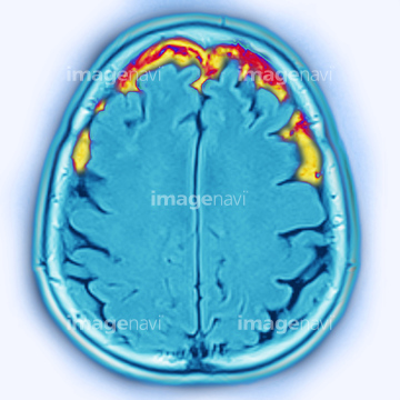
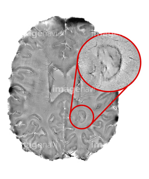
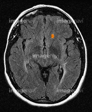
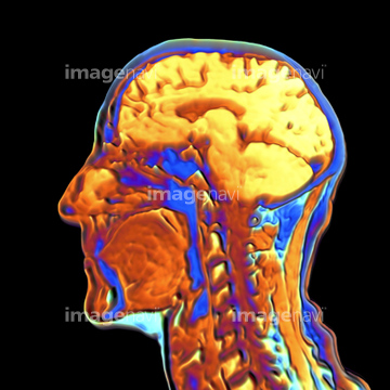
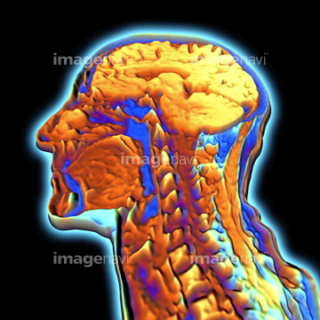
この検索結果には、Eclampsia, MRI scan、Brain, MRI scan、Osteoporosis on lumbar vertebrae.、Close-up of three injections、Brain scan、Brain, 3-D MRI scanなどが含まれています。
53156759
53156750
53156751
53156752
53156753
53156754
53156755
53156756
53156757
53156758
53156760
53156761
53156796
53156797

53156798
53156799
53156800
53156801
53156802
53156803
53156804
53156805
53156807
53156810
53156811
53156812
53156813
53156814
53156774
53156775
53156776
53156777
53156656
53156657
53156658
53156659
53156633
53156634
53156635
53156636
64021445
64021446
64021448
64021450
64021454
64021456
64071706
64071925
64071926
20562166
53156914
53156915
53156916
53156917
16918316
64021453
64021455
64021468
64021469
64021470
64021471
64021472
64042984
64059785
64059786
64049873
64071707
64071927
64021447
64021452
64021473
64064694
64066835
64049886
17260856
16900217
64022438
64022439
64022440
53156548
53156549
53156556
53156557
53156558
53156559
53156560
53156561
53156562

53156563
16918270
16918314
16918315
16918323
16918325
16918332
16918335
10941464
30072385
17201404
64021443
64021451
64043131
64040391
64040392
64056052
17218191
64110133
64021444
64064693
53156605
53156606
64066833
64066834
16932651
16932652
53156795
53156806
53156808
53156809
16918309
64110145
64060675
64062201
64062202
64062203
64188513
64188514
53156766
53156767
53156768
53156769
17218192
17218197
17218200
17218204

17218210
64021478
64219589
30342237
17247388
17201401
64021449
66000180
66000580
64021475
64021477
16918273
16918313
16918340
64071942
64051975
64172495
64172496
64161164
64161165
64161171
64161172
64161174
64161176
64161177
64161178
64161182
64161187
64161200
64161201
64161204
17235936
17235937
| 次ページ |
