HOME > 写真 > 医療・福祉 > 医療 > 手術
10,000件の写真素材が検索されました。
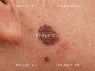
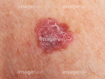
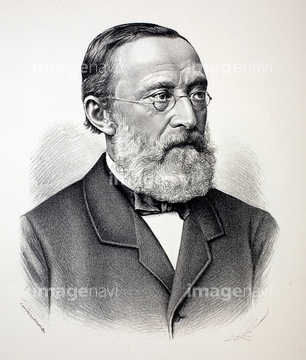
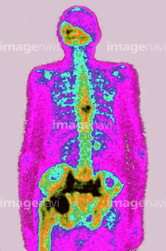
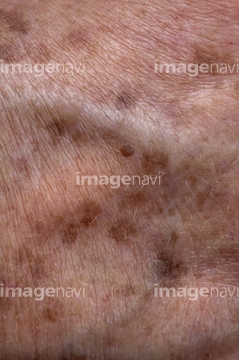
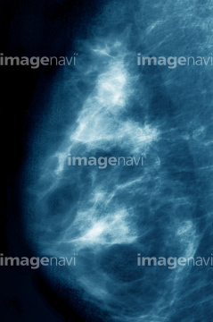
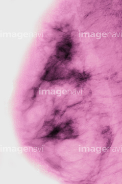
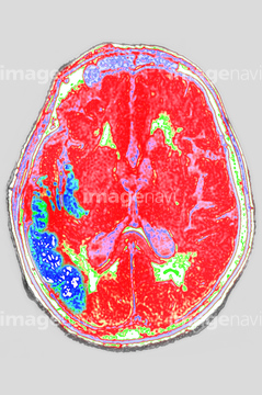
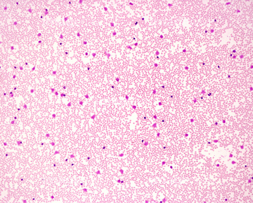
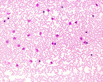
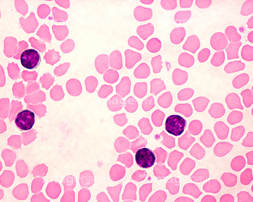
この検索結果には、1870 Rudolf Virchow German Scientist、Microscope use、Paget's disease.、Age spots on elderly skin、Breast cancer.、Clostridium botulinum.などが含まれています。
53156870
53156860
53156857
53156858
53156859
53156861
53156839
53156847
53156856

53156862
53156868
53156869

53156872
53156831
53156840
53156841
53156843
53156846
53156854
53156863
53156871
53156842
53156852
53156864
53156865
53156866
53156867
53156850
53156853
53156855
53156848
53156844
53156845
53156849
17239622
17239624
53156613
53156614
53156615
53156616
53156653
53156654
53156655
17239621
17239623
17239625
53136137
53156851

64118378
64118493
64223193
53156918
53156919
53156920
53156921
20523449
53156625
53156626
53156627
53156628
53156621
53156622
53156623
53156624
64058066
53156796
53156797

53156798
53156799
53156800
53156801
53156802
53156803
53156804
53156805
53156807
53156810
53156811
53156812
53156813
53156814
20523447
20523448
53156726
64222900
53156617
53156618

53156619
53156620

17238393
19967312
53156721
53156722
53156723
53156724
53156725
53156762
53156763
53156764
53156765
53156605
53156606
53156730
53156734
53156735
53156736
53156737
53156738
53156739
53156740
53156746
53156747
53156748
53156749
53156770
53156771
53156772
53156773
53156774
53156775
53156776
53156777
19967310
19967313
19967314
64048771
53156750
53156751
53156752
53156753
53156754
53156755
53156756
53156757
53156758
53156759
53156760
53156761

29253149
53156608
53156609
53156610
53156611
53156629
53156630
53156631
53156632
53157393
53157394
53157395
53157396
64210357
64066473
21591177
64147434
17205314
20568162
64194355
64044047
64139123
19967311
19967293
53156548
53156549
53156556
53156557
53156558
53156559
53156560
53156561
53156562

53156563
53156604
53156612
53156633
53156634
53156635
53156636
53156637
53156638
| 次ページ |
