HOME > ژتگ^ > ژY‹ئپEٹآ‹«–â‘è > ƒTپ[ƒrƒX‹ئ > ˆم—أپE•ںژƒ‹ئ
10,000Œڈ‚جژتگ^‘fچق‚ھŒںچُ‚³‚ê‚ـ‚µ‚½پB
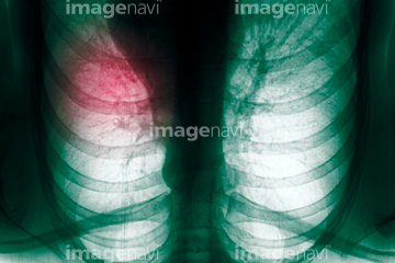
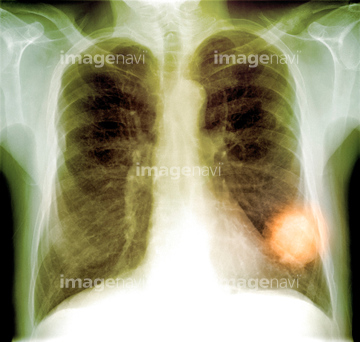
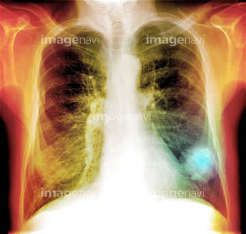
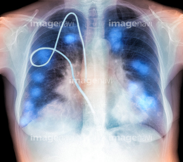
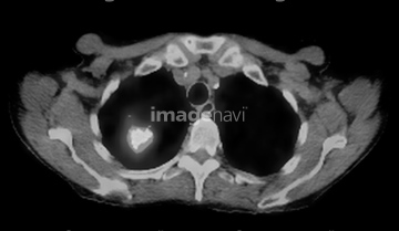
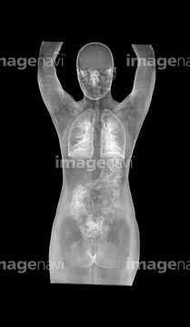
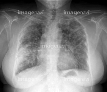
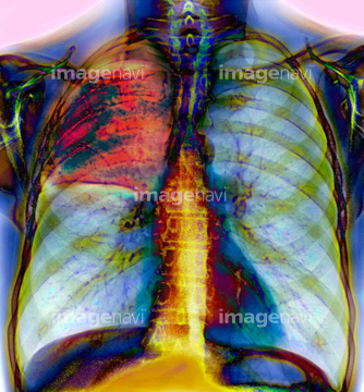
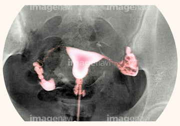
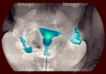
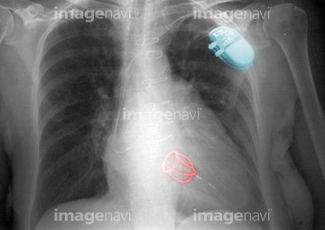
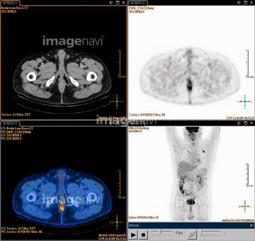
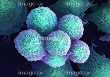
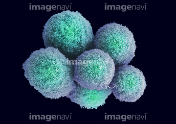
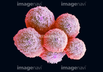
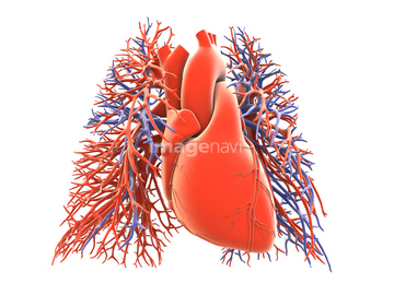
‚±‚جŒںچُŒ‹‰ت‚ة‚حپALung cancer, CT and PET scansپAFemale body, CT scanپALung disease, chest X-rayپAPneumonia, X-rayپA'Pneumothorax, X-ray'پA'Pneumothorax treatment, X-ray'‚ب‚ا‚ھٹـ‚ـ‚ê‚ؤ‚¢‚ـ‚·پB
54003423
64054760
64054761
64054762
64054763
53157381

53157382
53157383
53157384
30080580
17200422
17200423
64018259
64021679
64021680
64018256
64018257
64018258
64064324
64064325
64064326
64064320
64021686
17200424
64064329
53157292
53157293
53157294
53157295
53157296
53157297

53157298
53157299
64012589
64022444
64045497
64045498
64021704
64021705
64056244

64064327
64064328
64022451
64056243
64018336
64018337
64012965
64012966
53156766
53156767
53156768
53156769
64021668
64014345
64014346
20543697
30342201
30053470
30053474
64084816
64021649
64021650
64012556
64012557
20556615
20556621
64064331
64021643
64021644
64038957
64038958
64038959
64049883
64021335
64021336
64021669
30342179
64021529
64021530
64021531
30342182
64056411
53157373
53157374
53157375
53157376
53157377
53157378
53157379
53157380
64076174
64021762
64021763
30333663
64159403
16900160
64037859
64132656
64132657
64132658
64132659
64132660
21565721
64045443
64045444
64045496
10307797
64049870
10919280
64087127
64013375
64062613
64062614
64062616
64062621
64021645
64021646
64256046
64256047

64256048
64256049
64256050
64063389
64063390
64225107
64075069
64078141
64258710
64258711
64094074
64219669
64211182
64063379
64063380
64063381
64056239
10307795
20582175
64021655
64021656
64021657
64063345
64021626
64021627
64021754
64063346
20543690
30349690
64021318
| ژںƒyپ[ƒW |
