HOME > 写真 > 科学・テクノロジー > 科学 > DNA・細胞
10,000件の写真素材が検索されました。
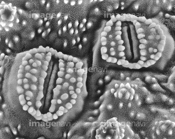
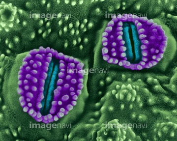
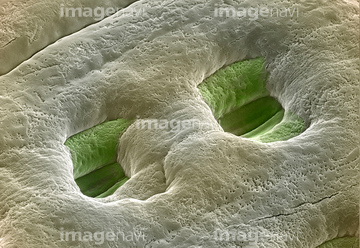
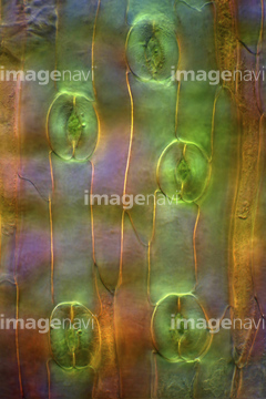
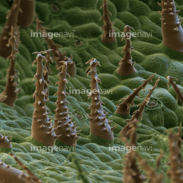
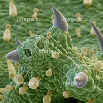
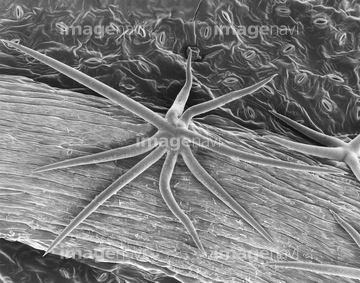
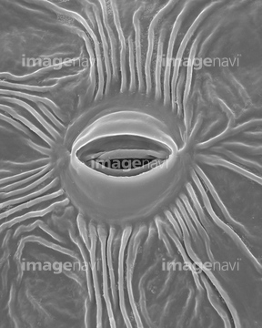
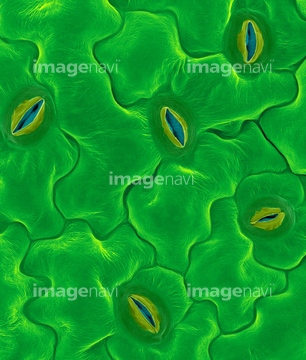
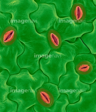
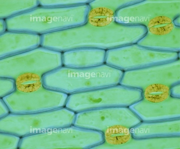
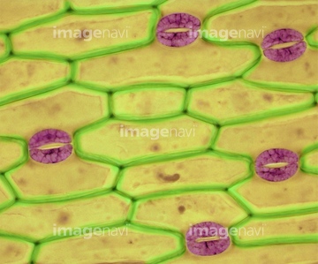
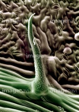
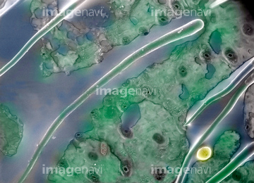
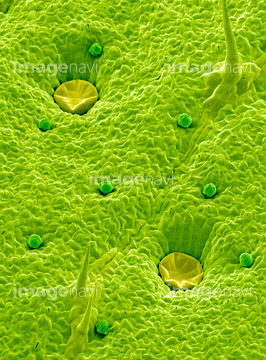
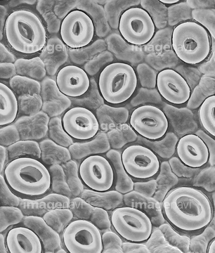
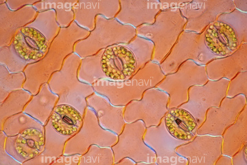
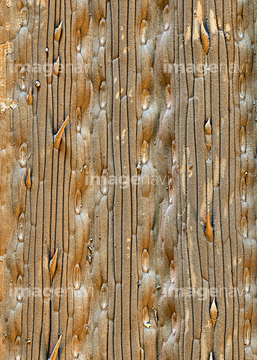
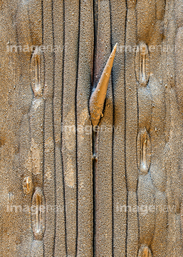
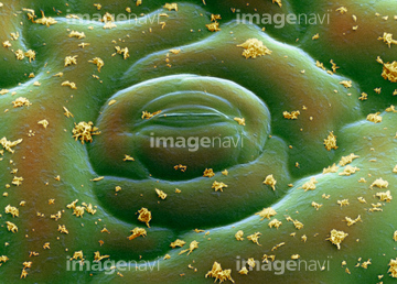
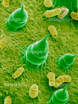
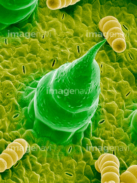
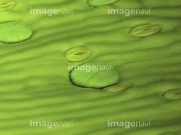
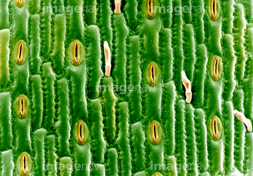
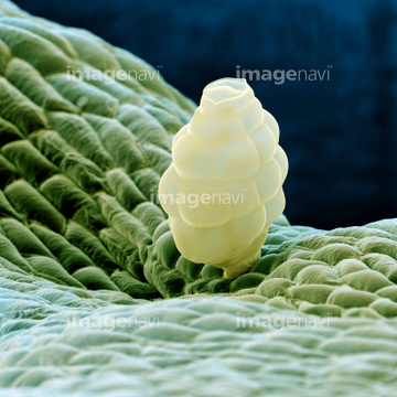
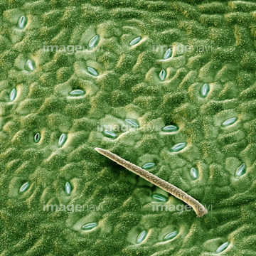
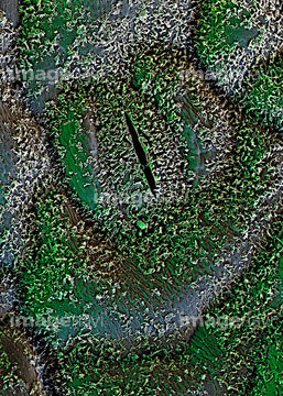
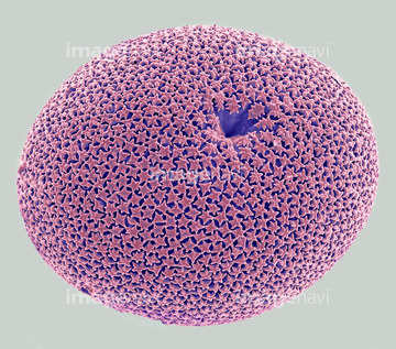
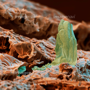
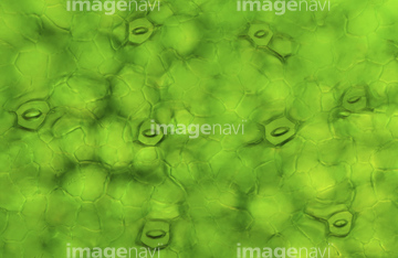
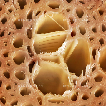
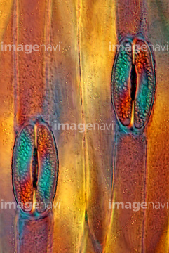
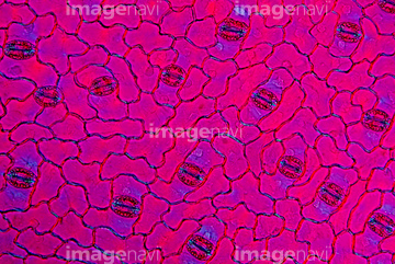
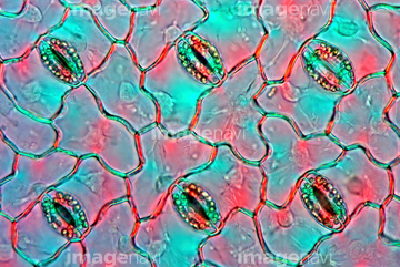
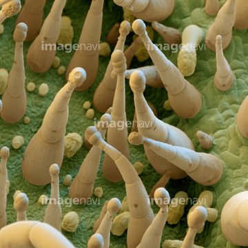
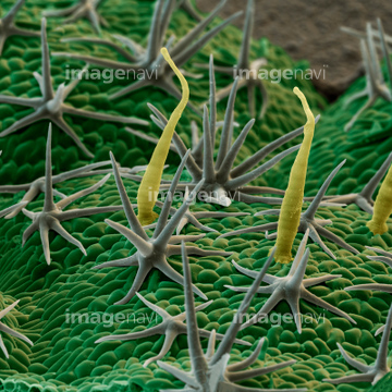
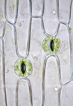
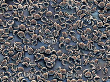
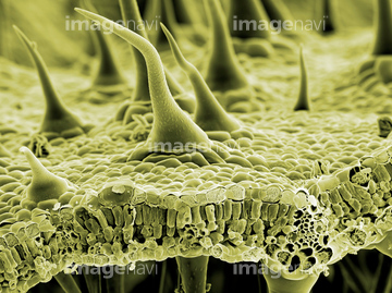
この検索結果には、Bottlebrush leaf surface, SEM、Oil glands on a leaf, SEM、Leaf stomata (Ohia Lehua tree; Metrosideros polymo…、Black Walnut tree (Juglans nigra)、Agave leaf、Plant leaf trichome (Hibiscus sp.), SEMなどが含まれています。
64005630
64005631
64005633
64005622

64004044
64005699
64005761
64005762
64005763
64208905
64208906
64208907
64005704
64005706
64004043
64005632
64005647
64255079
64255080
64210424
64005688
64151007
64005705
64089520
64089521
64005629
64005638
64089438
64089507
64137288
64137289
64137290
64137291
64145495
64145512
64155055
64005646
64005675
64005689
64005765
64005766
64005709
64114726
64005643
64005641
64005734
64005735
64005683
64005684
64157866
64056494
64005642
64005644
64005648
64005657
64005665
64005666

64089505
64089506
64090375
64090376
64199842
64005390
64150985
64137210
64005681
64005682
64089396
64089397
64089398
64005722
64005627
64005628
64005637
64089408
64006296
64089522
64089523
64005703
64005716
64005717
64148624
64224866
64006294
64006295
64148626
64098748
64098749
64099743
64105240
64105241
64105251
64105268
64105281
64005691
64042130
64042146
64042131
64042147
64089399

64114192
64029676
64195202
64195203
64195206
64195208
64213976
64077317
64259882
64259884
64259885
64259887
64259888
64259889
64259890
64259891
64005718
64064827
64061076
64061077
64061078
64061079
64061080
64061081
64061082
64005640
64005658
64255901
64008579
64098756
64105252
64105253
64005635
64005636
64064830
64041764
64076418
64005661

64061093
64061094
64061095
64061114
64012433
64005663

64195204
64195205
64056945
64012326
64078803
64056946
64040850
| 次ページ |
