HOME > 写真 > 科学・テクノロジー > 科学 > DNA・細胞
10,000件の写真素材が検索されました。
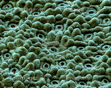
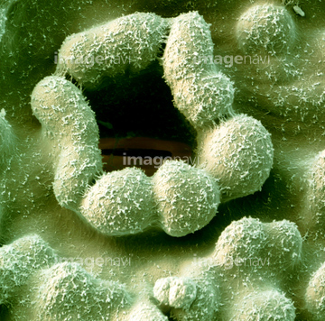
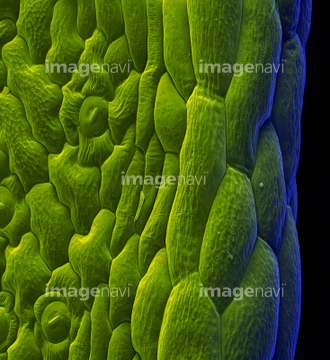
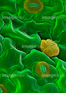
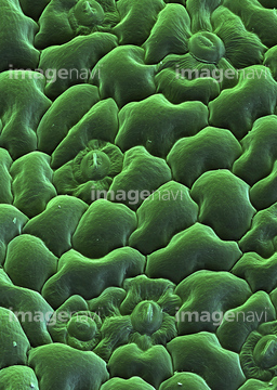
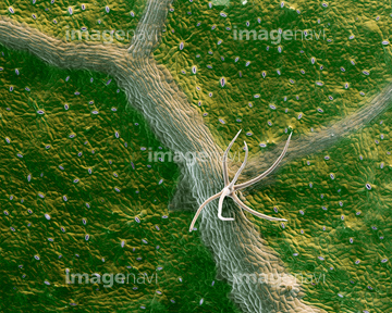
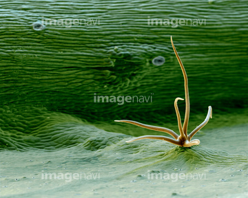
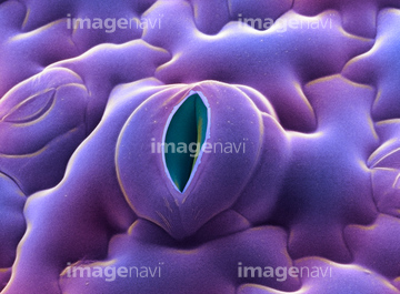
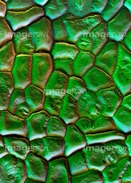
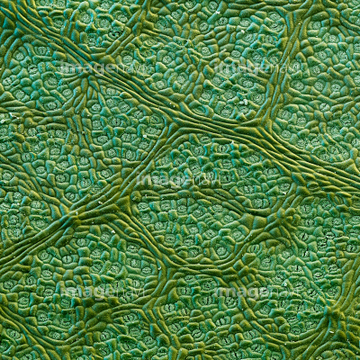
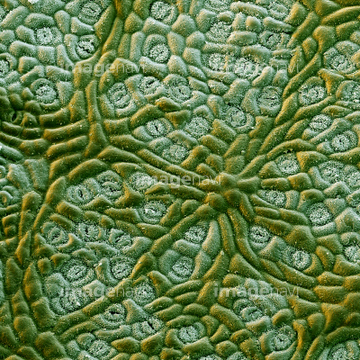
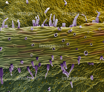
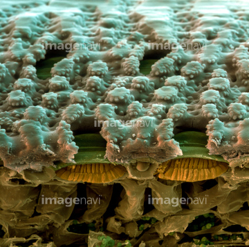
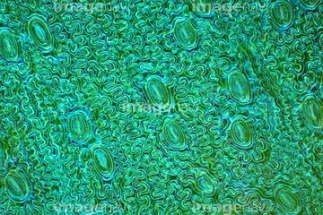
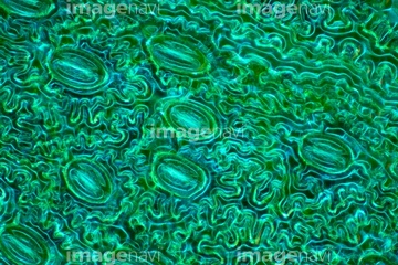
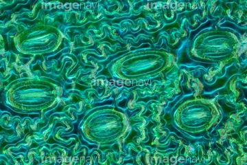
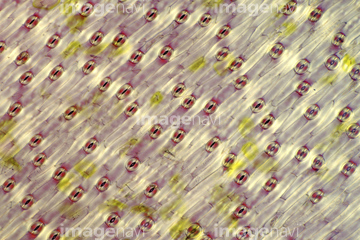
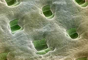
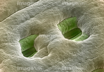
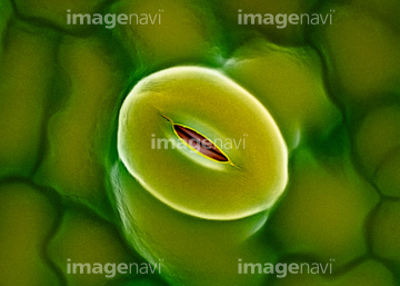
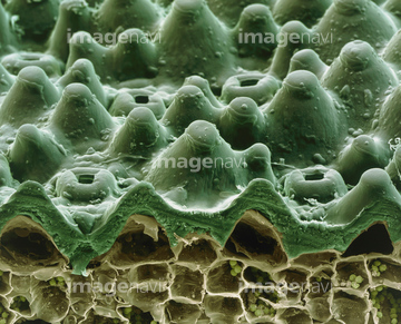
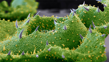
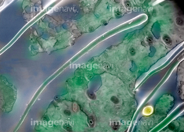
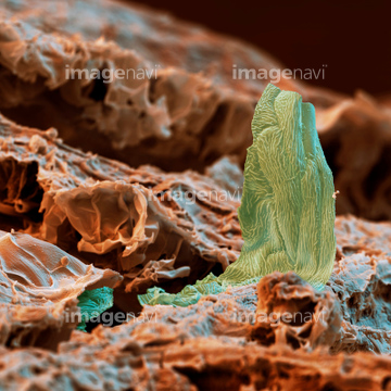
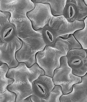
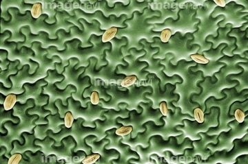
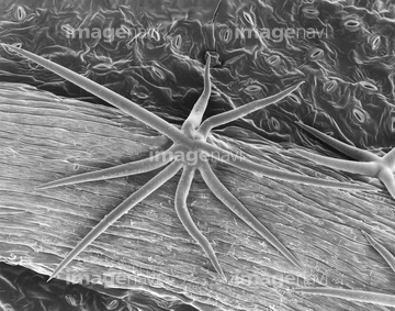
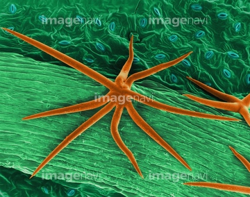
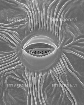
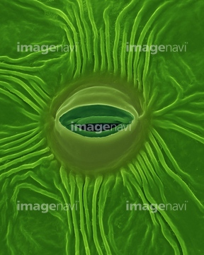
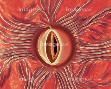
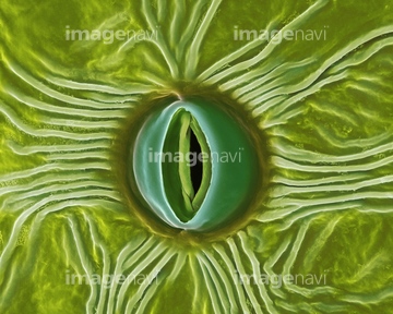
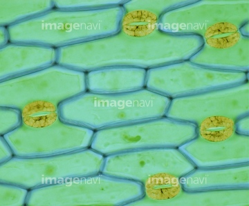
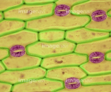
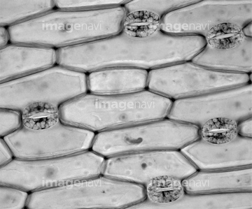
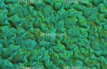
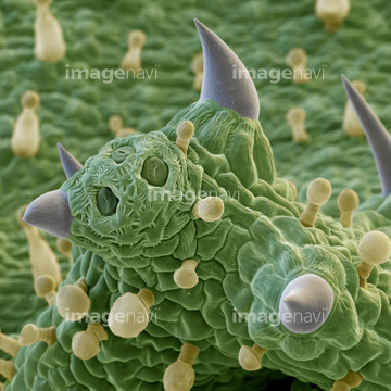
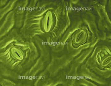
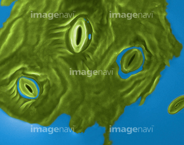
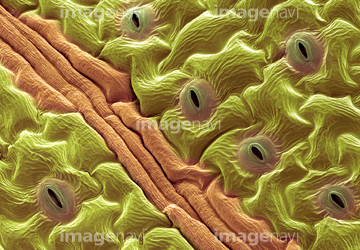
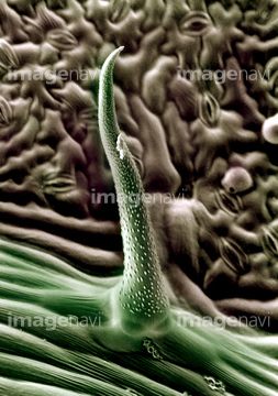
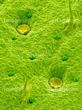
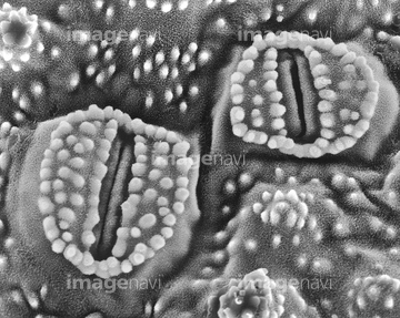
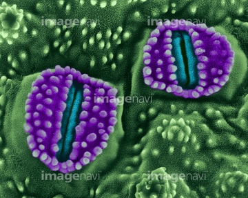
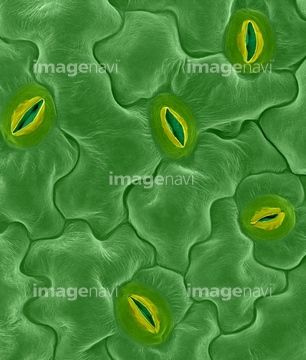
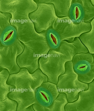
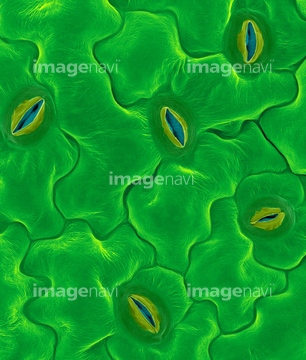
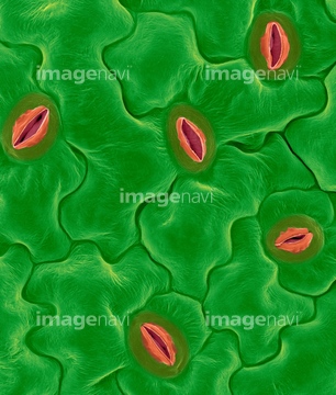
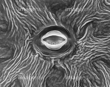
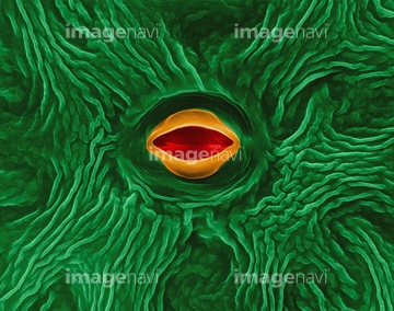
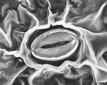
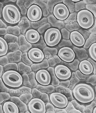
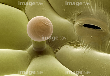
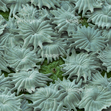
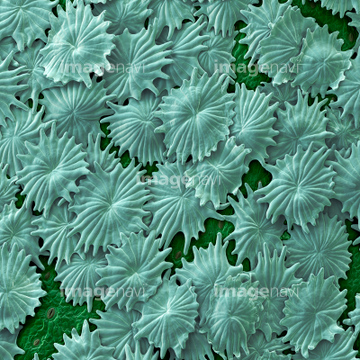
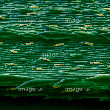
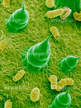
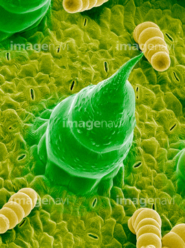
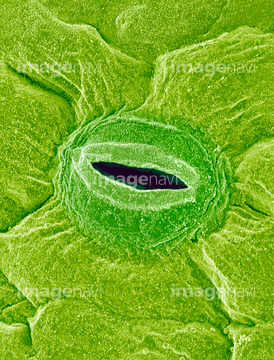
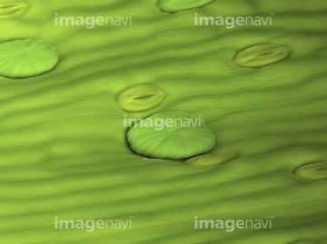
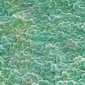
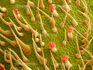
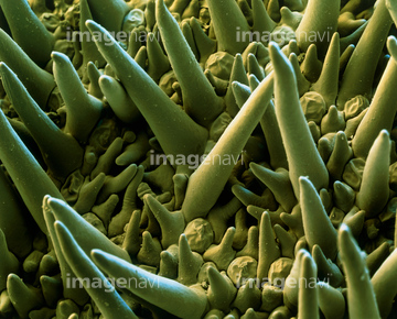
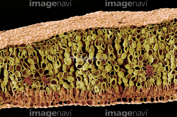
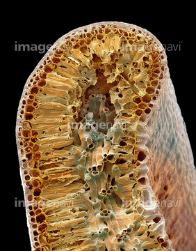
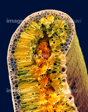
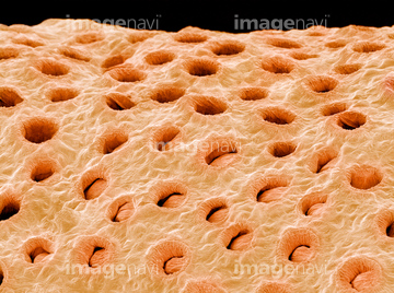
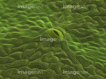
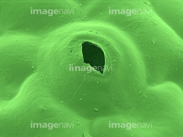
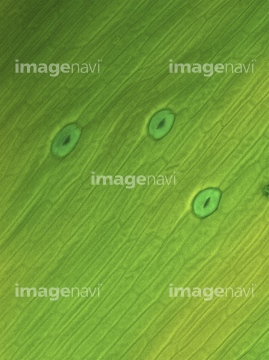
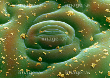
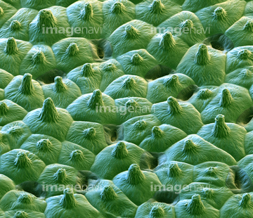
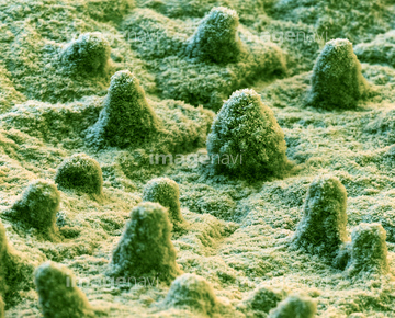
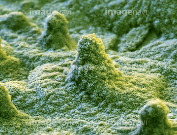
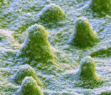
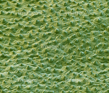
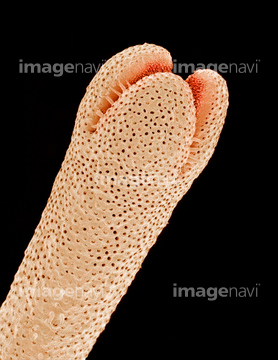
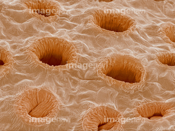
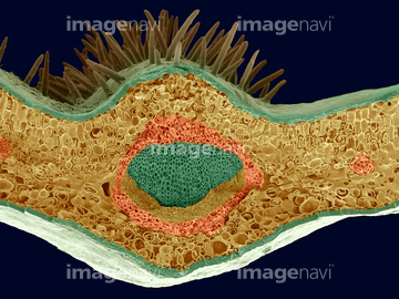
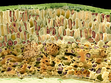
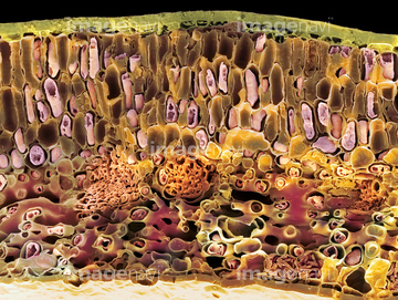
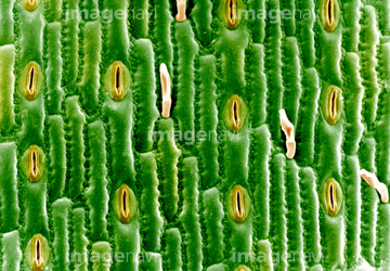
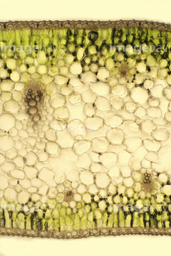
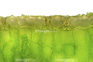
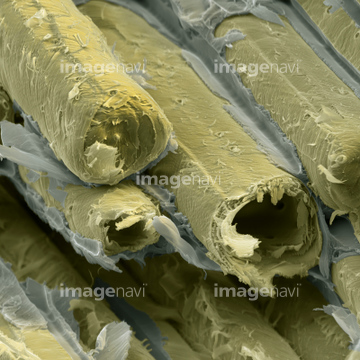
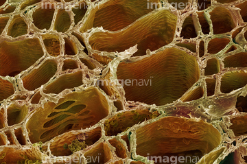
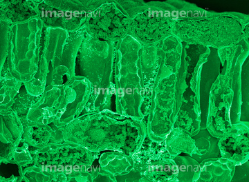
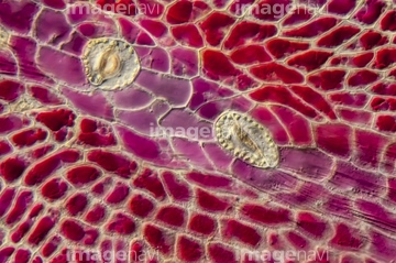
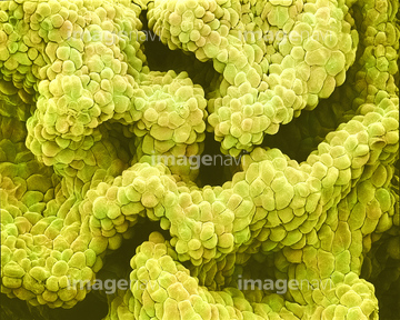
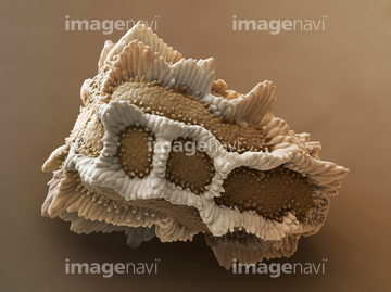
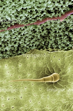
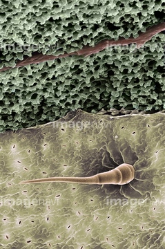
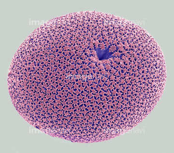
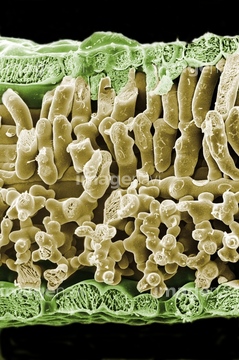
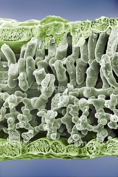
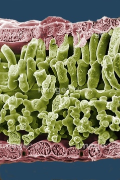
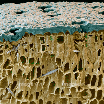
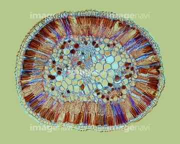
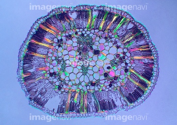
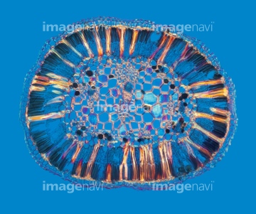
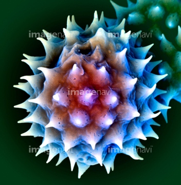
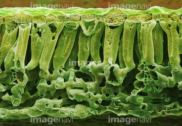
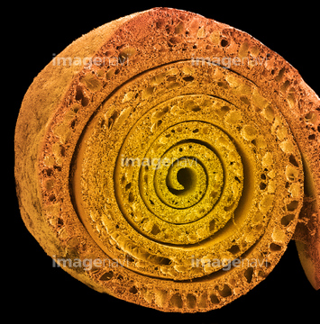
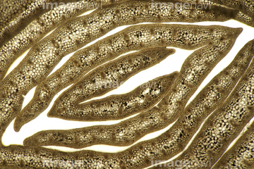
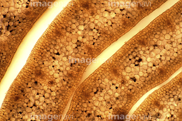
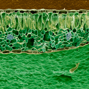
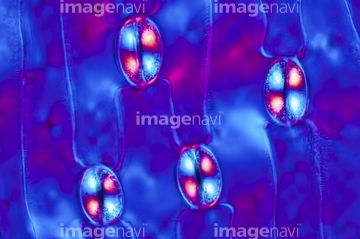
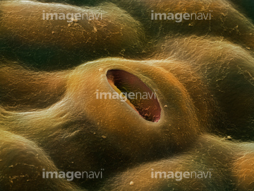
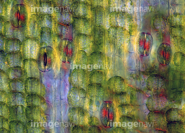
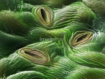
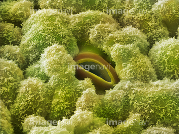
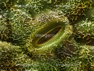
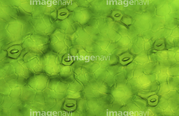
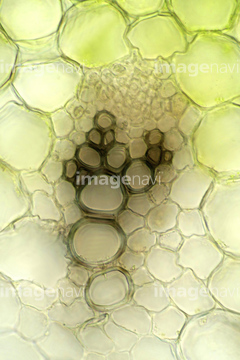
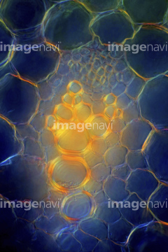
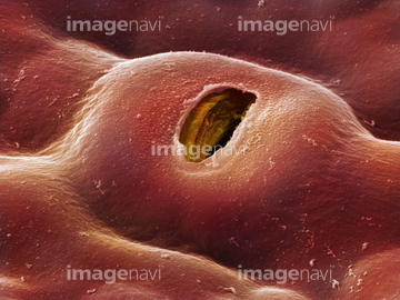
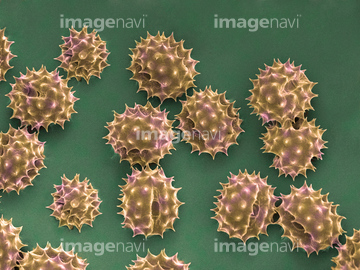
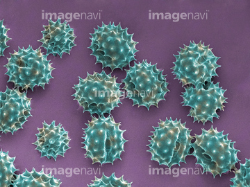
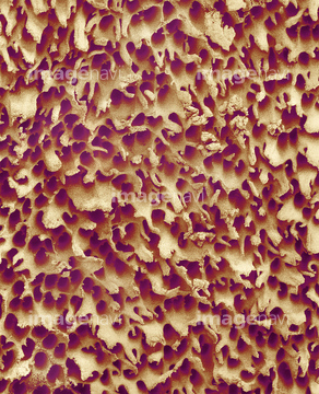
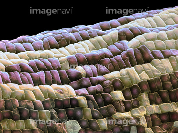
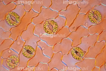
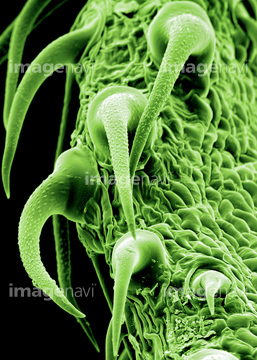
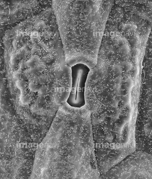
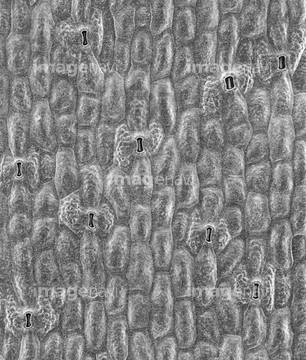
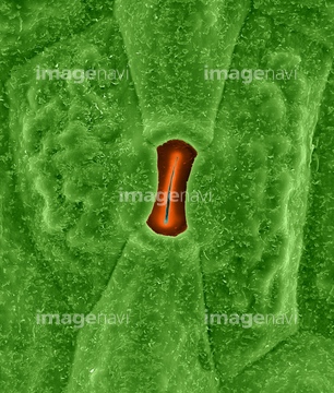
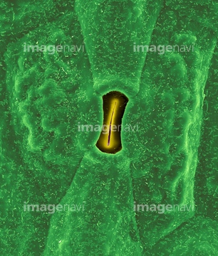
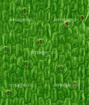
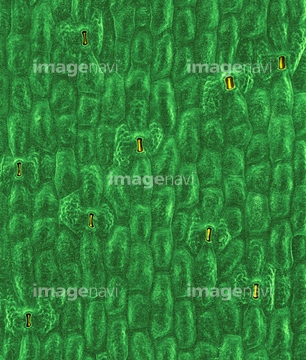
この検索結果には、Monilaria leaf、Agave leaf、Kleinia leaf、Rosemary leaf、Plant stomata, LM、Plant stomata, light micrographなどが含まれています。
64005699
64005622
64005630
64005631
64005633
64005761
64005762
64005763
64005683
64005684

64004044
64005706
64089520
64005681
64005682
64005675
64004043
64208905
64208906
64208907
64089521
64005704

64255079
64255080
64005646
64005647
64005705
64005641
64005688
64005766
64005658
64151007
64210424
64089438
64089505
64089506
64089507
64199842
64089522
64005642
64005643
64005644
64005648
64145495
64145512
64155055
64005638
64005665
64005666
64005689
64005765
64005709
64137288
64137289
64137290
64137291
64150985
64137210
64005687
64090375
64090376
64005734
64005735
64089523
64114726
64005629
64005632
64005657
64005627
64005628
64005637
64005716
64005717
64005722
64148624
64005718
64005390
64005690
64005726
64005727
64006296
64157866
64056494
64148626
64005703
64042796
64042797
64042798
64042800
64042801
64042803
64042804
64042805
64042806
64089396
64089397
64089398
64089399

64009115
64009116
64009118
64006294
64006295
64004133
64004142
64004143
64224866
64195202
64195203

64195204
64195205
64213976
64077318
64077442
64077443
64089408
64005344
64005411
64005420
64005423
64009025
64042130
64042146
64042147
64162969
64006239
64006660
64029676
64005640
64106440
64064827
64005691
64195206
64195208
64005663
64213957
64087657
64087662
64255901
64006419
64006420
64003460
64012659
64012432
| 次ページ |
