HOME > 写真 > 科学・テクノロジー > 科学 > DNA・細胞
10,000件の写真素材が検索されました。
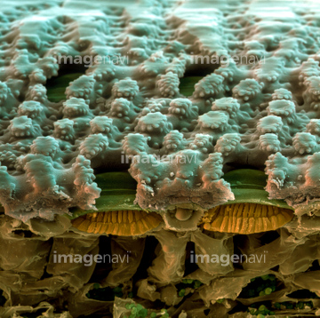
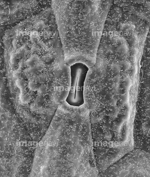
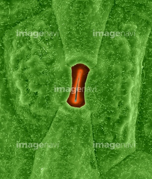
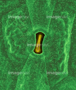
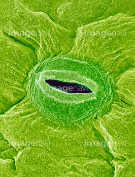
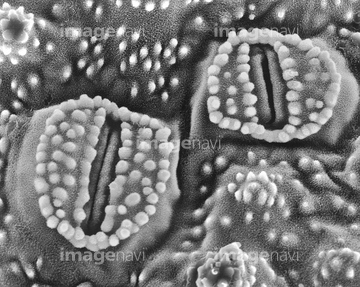
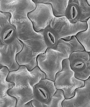
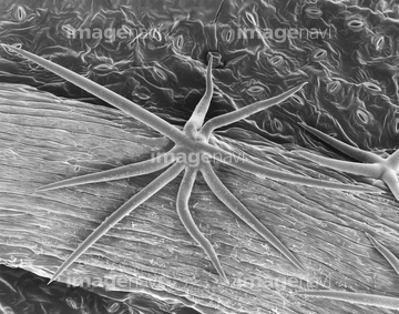
この検索結果には、Prickly pear cactus (Opuntia spp.) stoma, SEM、Sunflower leaf, SEM、Leaf pore, SEM、Stomata of scouring rush (Equisetum sp.), SEM、Leaf stomata (Arabidopsis sp.), SEM、Plant leaf trichome (Hibiscus sp.), SEMなどが含まれています。
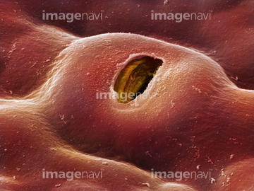
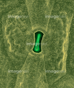
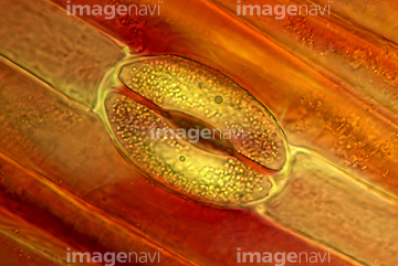
64005705
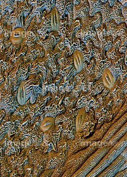
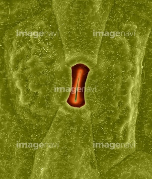
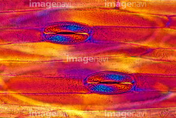
64005706
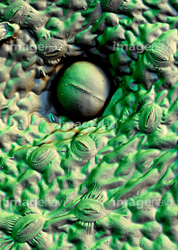
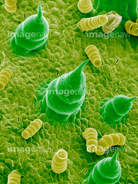
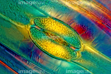
64005704
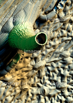
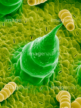
64005703
64089521
64087657
64089520
64005707
64005761
64005762
64005763
64087662
64089438
64089505
64089506
64089507
64199842
64005633
20542488
64089399
64150985
64137210
64145495
64145512
64155055
64208905
64208906
64208907
64210424
64005646
64005647
64089522
64089523
64004043
64005716
64005717
64005722
64151007
64137288
64137289
64137290
64137291
64005622
64005630
64005631
64005699
64224866
64089396
64089397
64089398
64090375
64090376
64005663
64162969
64089408
64005718

64004044
64259886
64098748
64098749
64099743
64105240
64105241
64105251
64105268
64105281
64006295
64006296
64087655
64089383
64089384
64259974
64005641
64098756
64105252
64105253
64255079
64255080
64005660
64005662
64005734
64005735
64056494
64259882
64259884
64259885
64259887
64259888
64259889
64259890
64259891
64099687
64105289
64105290
64105291
64128977

64003747
64005390
64250245

64005683
64005684
64005688
64157866
64176956
64259966
64259968
64259969
64259973
64005632
64005637
64005665
64005681
64005682
64005709
64003785
64002863
20530800
20542520
20542521
64259972
64005719
64005720
64005737
64148624
64114726
64005627
64005628
64005629
64005638
64005642
64005643
64005644
64005648
64005657
| 次ページ |
