HOME > 写真 > 花・植物 > その他植物 > ハーブ
10,000件の写真素材が検索されました。
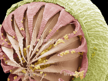
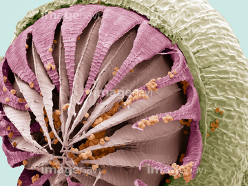
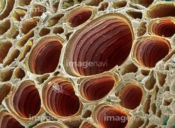
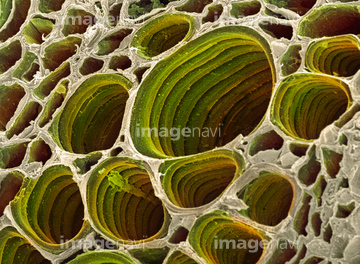
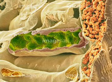
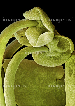
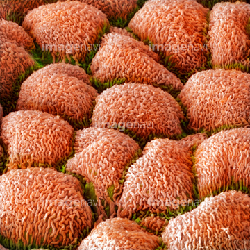
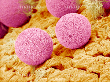
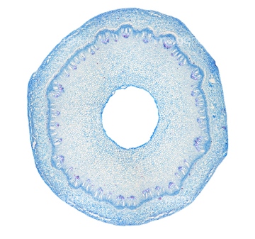
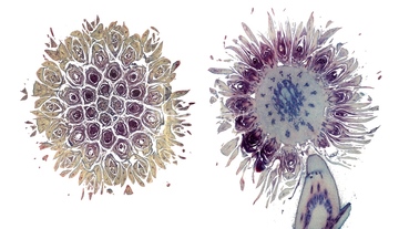
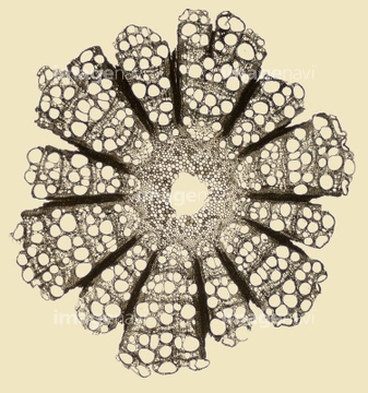
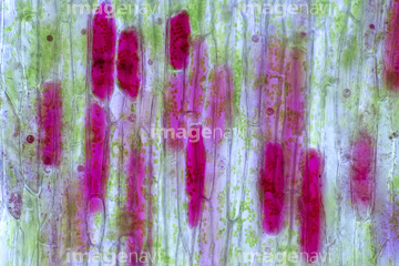
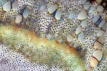
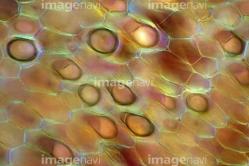
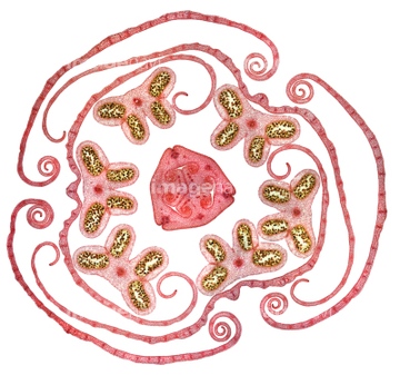
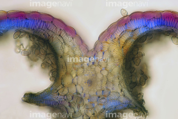
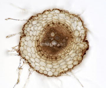
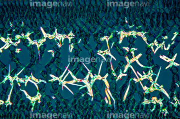
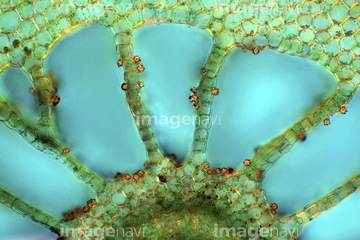
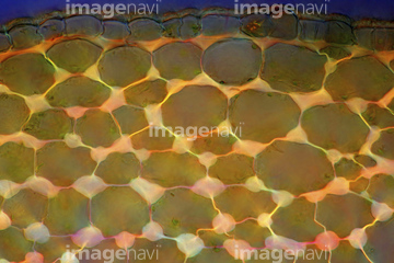
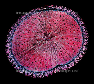
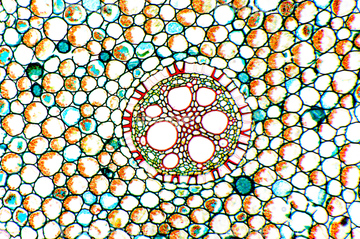
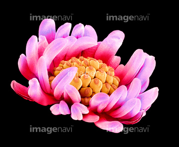
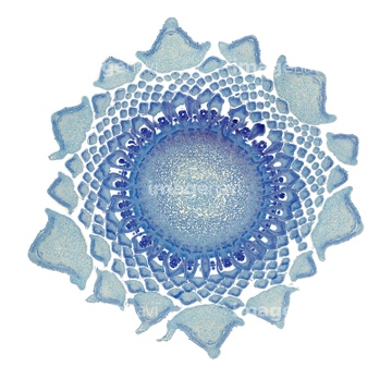
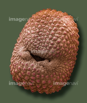
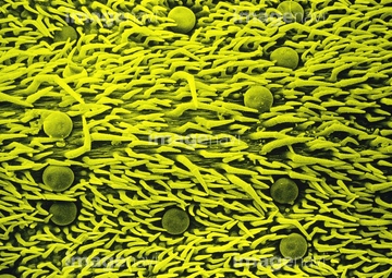
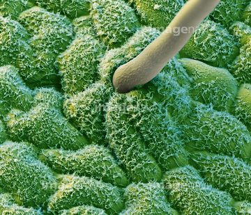
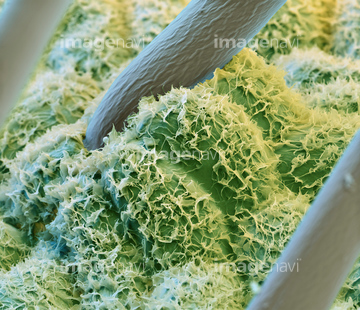
この検索結果には、Xylem plant cells, SEM、Walnut wall, SEM、Disc floret, SEM、Comfrey flower petal, SEM、Chrysanthemum root, light micrograph、Buttercup flower, SEMなどが含まれています。

64006171
64006172
64006173
64005746
64005747
64005745
64006162
64006163
64005441
64006169
64006170
64006160
64006161
64006167
64006168
64041764
64006159
64005744
64005759
64062364
64008579
64006166
64005738
64005739
64005748
64005749
64005750
64005751
64005752
64005754
64005755
64006308
64006307
64005740
64005741
64005442
64005753
64006164
64006165
64013180
64114731
64078803
64012659
64064830
64008774
64008775
64009113
64009114
64009116
64074760
64085181
64085182
64006309
64006154
64006155
64212520
64212522
64212523
64212524
64212525
64006310
64006311
64233701
64006145
64012389
64012390
64012461
64012462
64012463
64012475
64012666
64008582
64009047
64009048
64009049

64009115
64009118
64006306
64010623
64006458
64006459
64006460
64171741
64006279
64006152
64006479
64006461
64006462
64006463
64006464
64006457
64075216
64005722
64147360
64147365
64147380
64006158
64005622
64005623
64005624
64117897
64006452
64006453
64006455
64006456
64078793
64012409
64009025
64117902
64195156
64195202
64195203
64195206
64195208
64261315
64261320
64261323
64261327
64261341
64261343
64261352
64261355
64261356
64261357
64261358
64261360
64212518
64247479
64233503
64040153
64006312
64147359
64147362
64147364
64147370
64140433
64255075
64073056
64212521
64111628
64111631

64013905
64049404
64049405
64005648
64005650
64005652
64005653
64005654
64005655
64005656
64005689
64006621
64006622
64006632
64006633
64008102
64008680
64008681
64042130
64042131
64042146
64042147
| 次ページ |
