HOME > 写真 > 科学・テクノロジー > 科学 > DNA・細胞
10,000件の写真素材が検索されました。
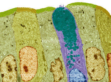
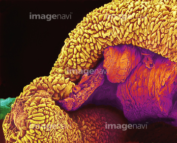
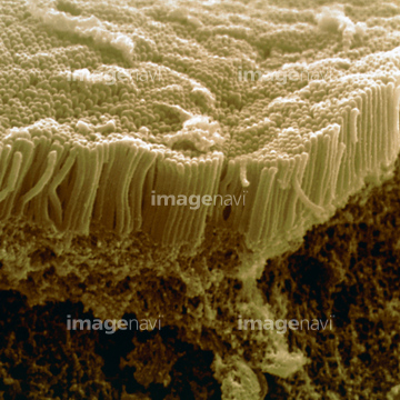
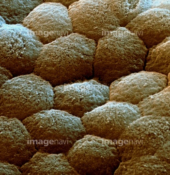
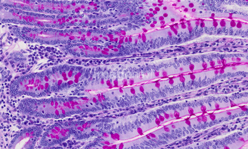
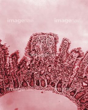
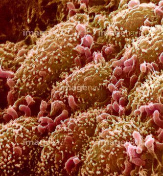
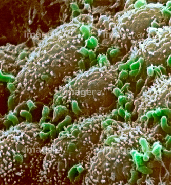
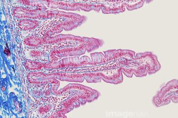
この検索結果には、Peyer's patch, light micrograph、Small bowel, light micrograph、Duodenum, light micrograph、Villus surface of the small intestine, SEM、Intestinal villi, SEM、Pancreas tissue, SEMなどが含まれています。
64008180

64225133
64075661
64021539
64021540
64021541
64021571
64049910
64206819
64008744
64008745
64073608
64077368
64021549
64021550
64021551
64061536
64247696
64088343
64049911
64021569
64021568
64021554
64021570
64196734
64073343
64021534
64021537
64060825
64060826
64074891
64251226
64196715
64197487
64197488
64090479
64090480
64090490
64213913
64213914
64213915
64152547
64076021

64225083
64009119
64009121
64145925
64021545
64008086
64166182
64055781
64021548
64021552
64056271
64021532
64021535
64021536
64021538
64021546
64008084
64008085
64008155
64008156
64008157
64077369
64260019
64073304
64251229
64002984
64002985
64008746
64008751
64021533
17202471
17202472
64073988
64076022
64076023
64166159
64021512
64021592
64060827
64060828
64008178
64073285
64090152
64055763
64224937
64209800
64072010
64072070
64072071
64076227
64076228
64021528
64040200
64060814

64060815
64060816
64009124
64009125
64009132
64009133
64009134
64009135
64009136
64009137
64021658
64021659
64009035
64009164

64009166
64009168
64197106
20542482
64199170
64199171
64098209
64114553
64073311
64021596
64021597
64021598
64116672
64106069
64106074
64106079
64106080
64106092
64057542
64060933
64066076
64071989
64072730
64072745
64072748
64072750
64073274
64073275
64073276
64073301
64073308
64085258
64196724
64196725
64213876
64172510
64172534
64090457
64090463
64214176
64166166
64234667
64234718

64225140
64076362
64076363
64005344
64005726
64005727
64008876
64076353
64076354
64076360
64076361
64077278

64003062
64003120
| 次ページ |
