HOME > 写真 > 科学・テクノロジー > 科学 > DNA・細胞
10,000件の写真素材が検索されました。
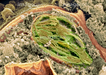
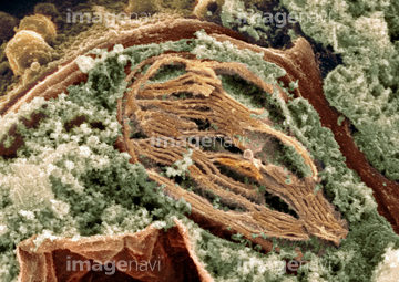
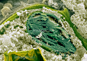
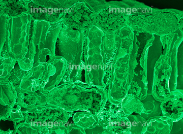
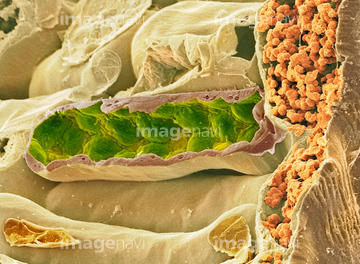
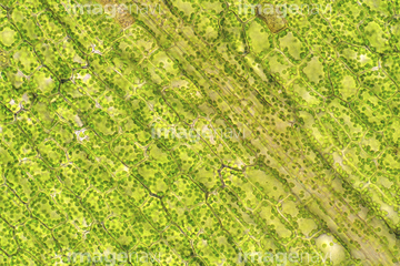
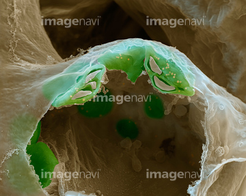
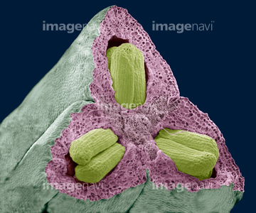
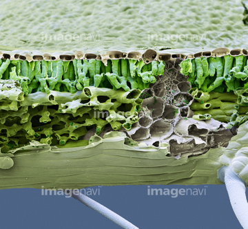
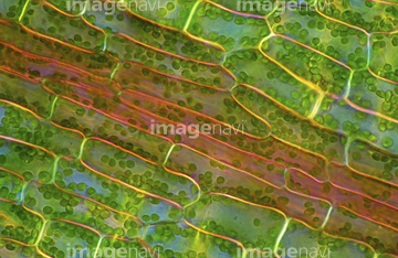
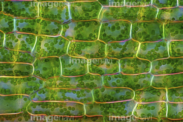
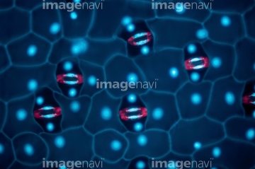
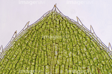
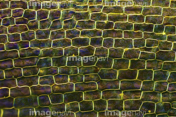
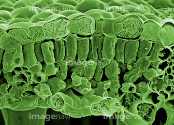
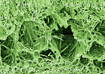
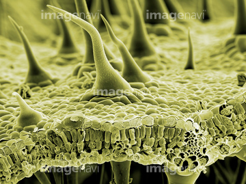
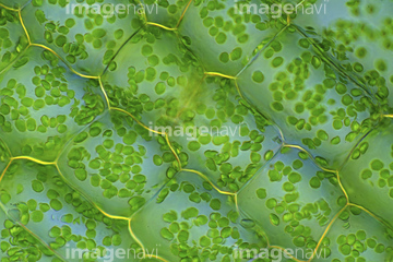
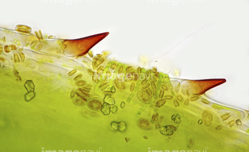
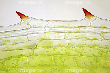
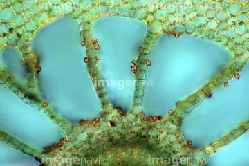
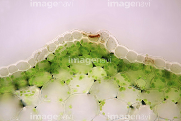
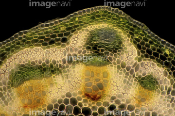
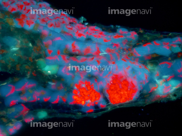
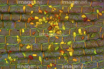
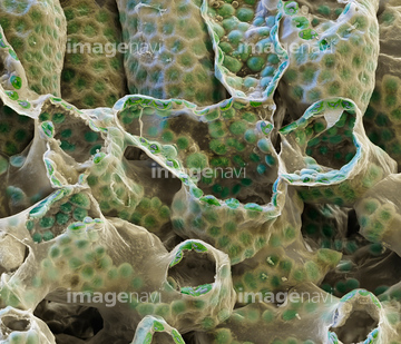
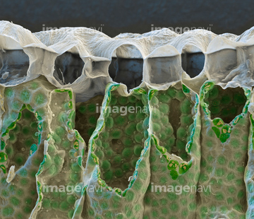
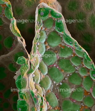
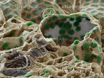
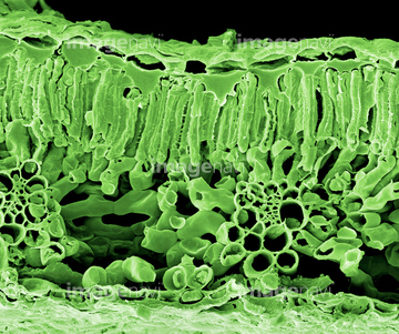
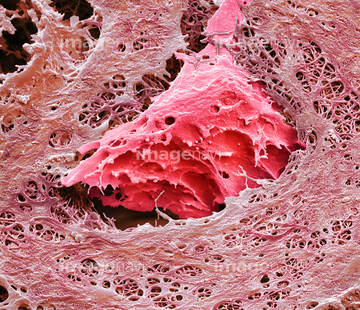
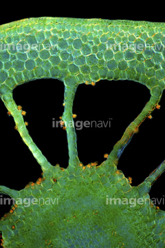
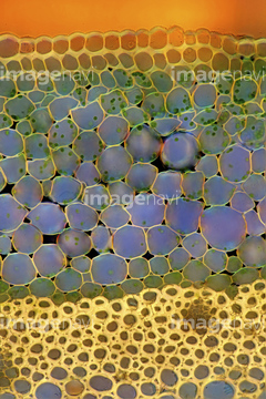
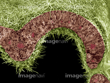
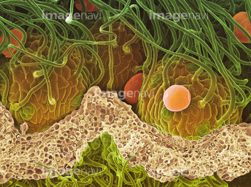
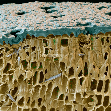
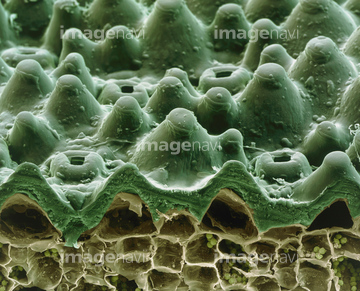
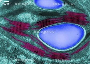
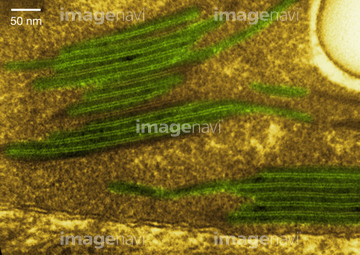
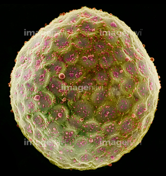
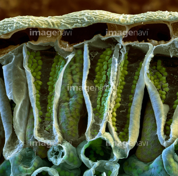
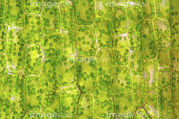
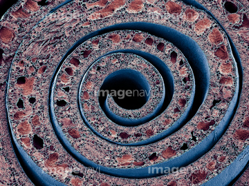
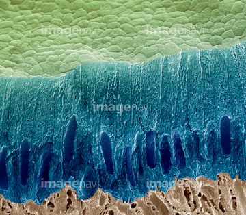
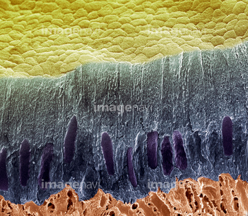
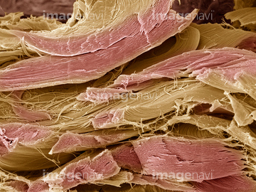
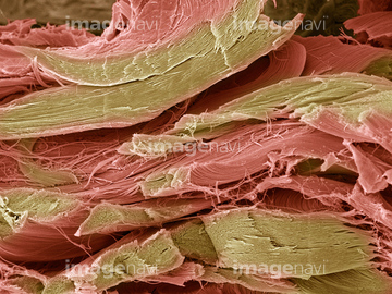
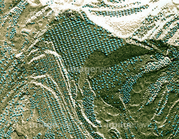
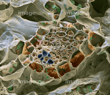
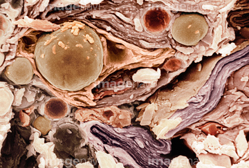
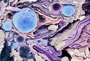
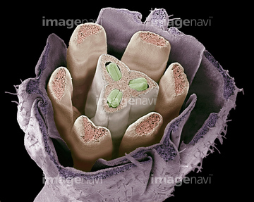
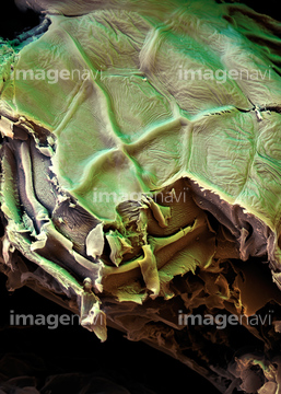
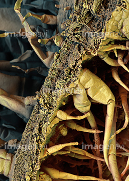
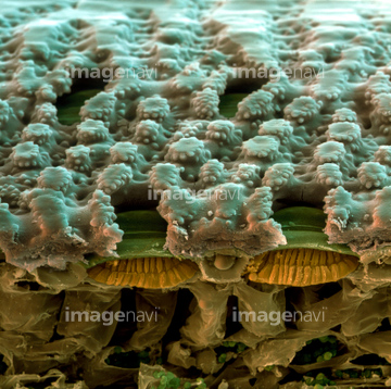
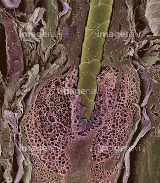
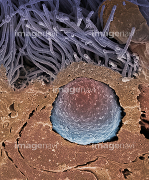
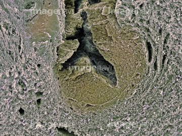
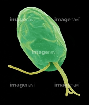
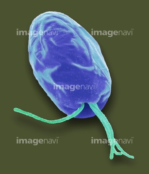
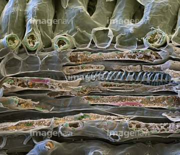
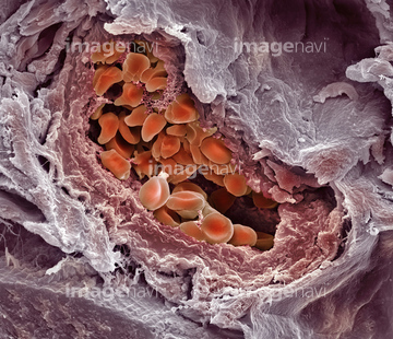
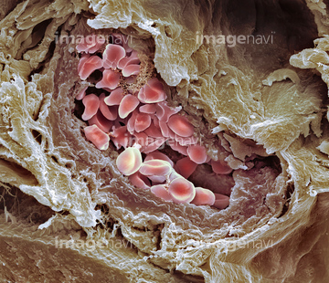
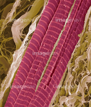
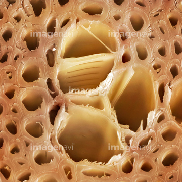
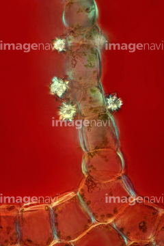
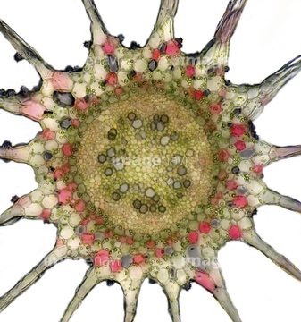
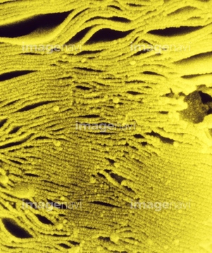
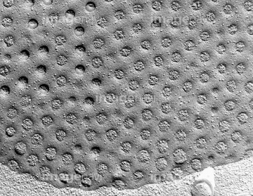
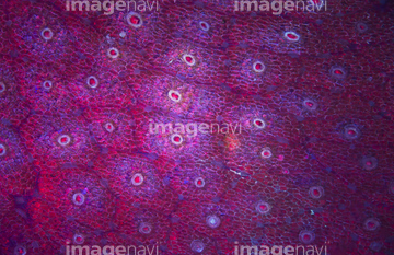
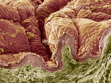
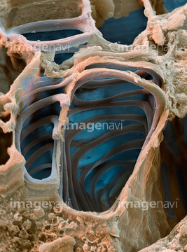
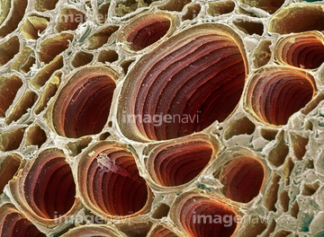
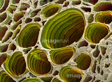
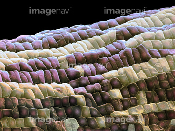
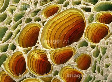
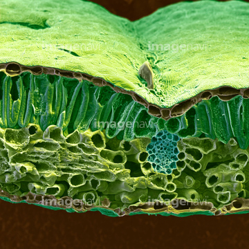
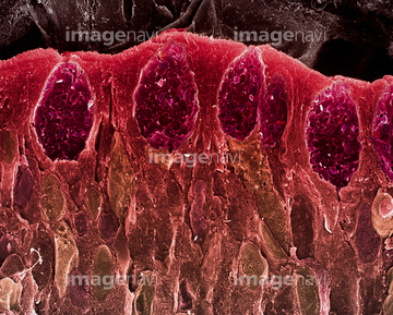
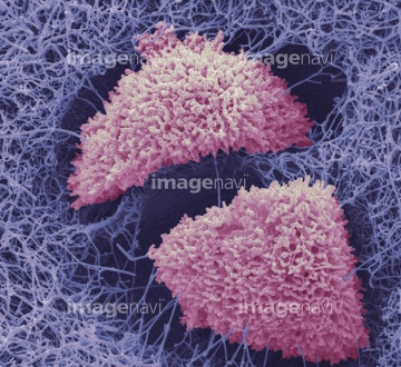
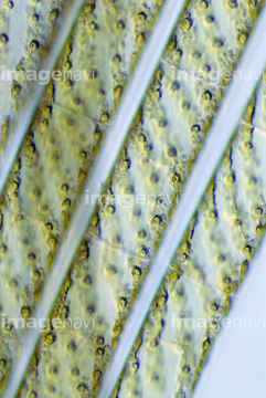
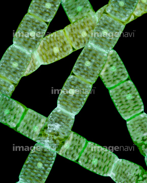
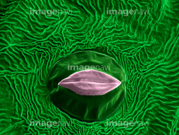
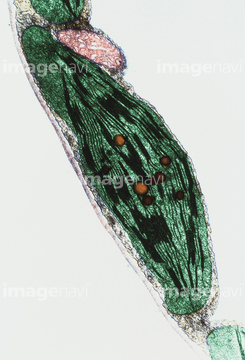
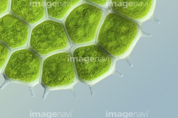
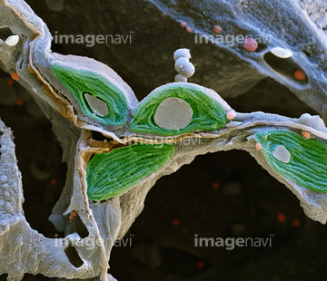
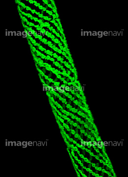
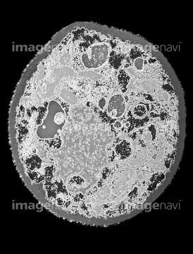
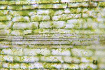
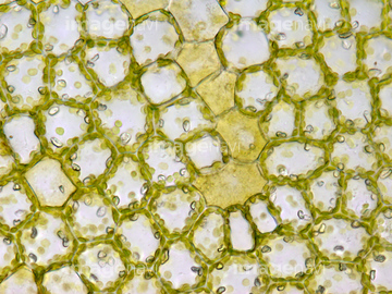
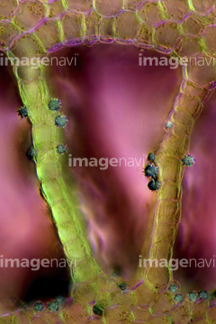
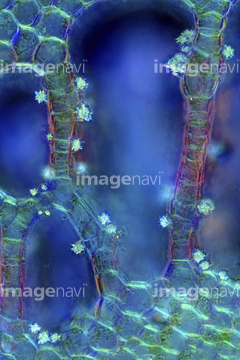
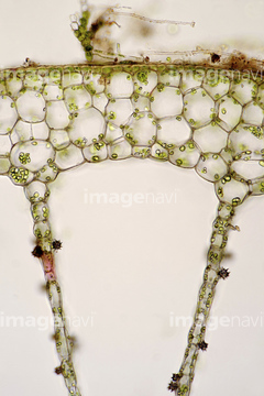
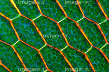
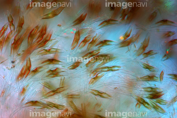
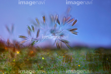
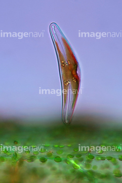
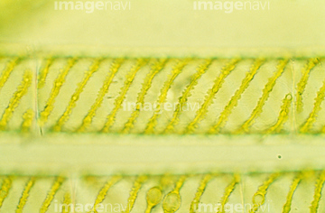
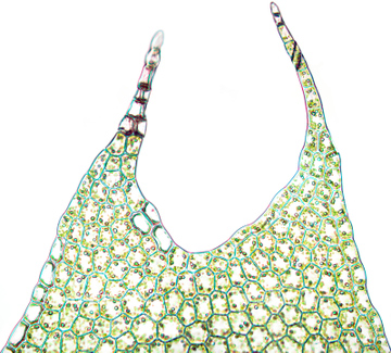
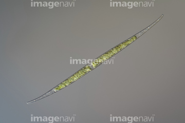
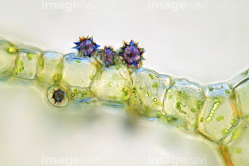
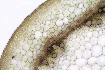
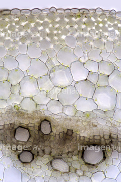
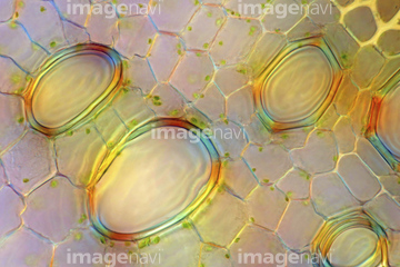
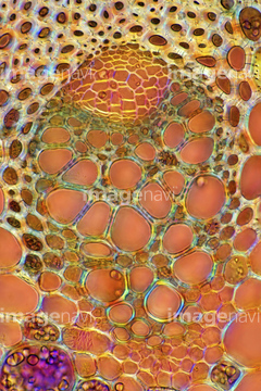
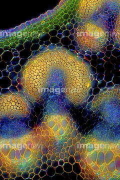
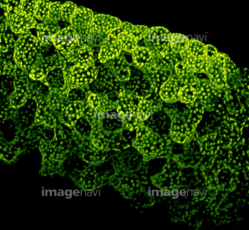
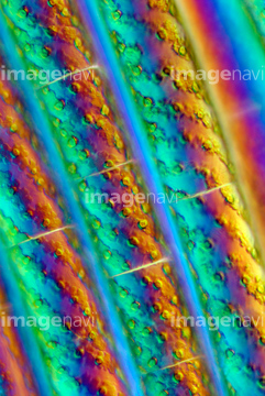
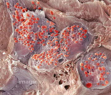
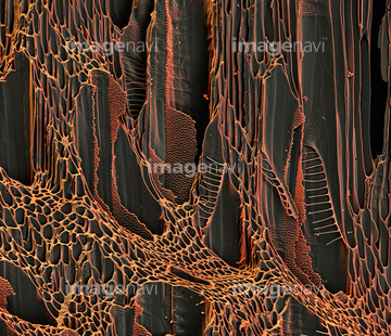
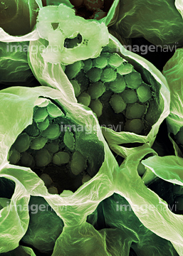
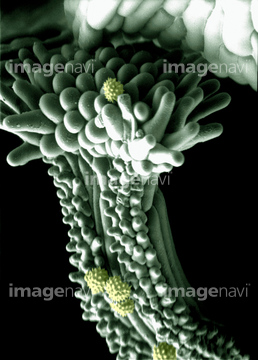
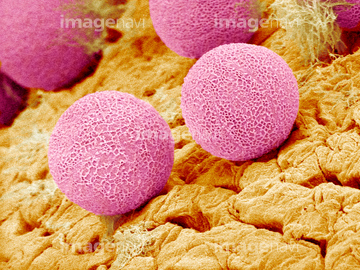
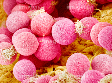
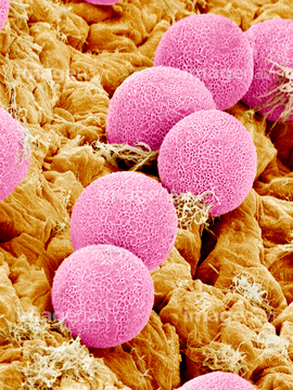
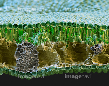
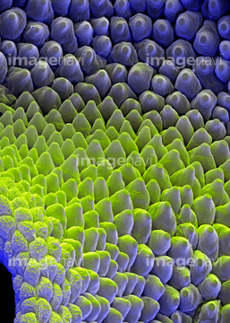
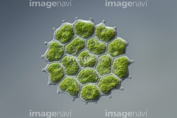
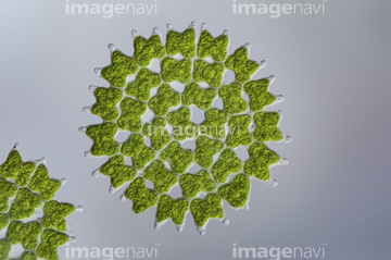
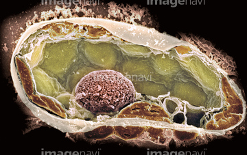
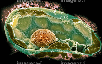
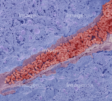
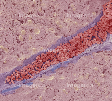
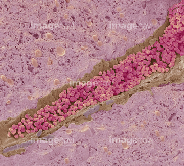
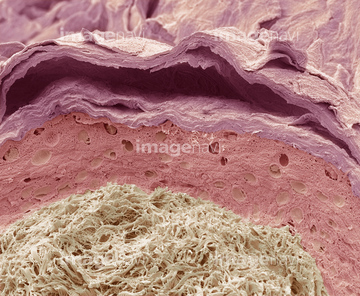
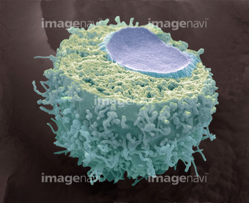
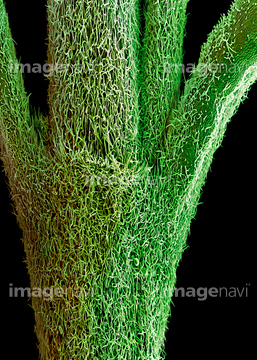
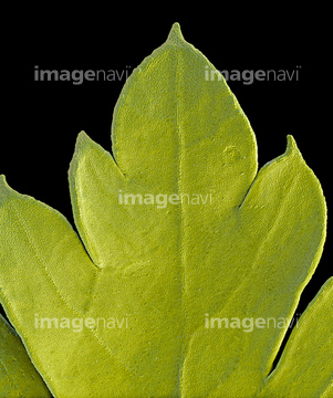
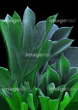
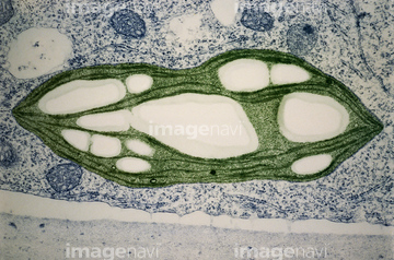
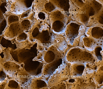
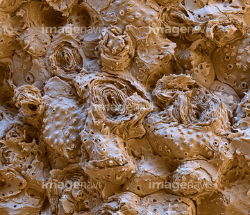
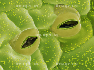
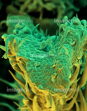
この検索結果には、Christmas rose leaf, SEM、Canadian pondweed (Elodea canadensis), light micro…、Coiled lily leaf, SEM、Hair follicle, SEM、Tooth enamel formation, SEM、Tendon fibres, SEMなどが含まれています。
64009047
64009048
64009049
64009025
64010742
64010743
64010744

64009115
64009116
64009118
64078782
64013180
64012409
64119747
64040850
64012431
64012433
64233503
64056946
64056947
64056948
64056949
64012403
64072558
64008102
64005713
64005640
64005641
64002854
64005660
64005701
64005702
64058508
64058509
64058510
64008084
64008085
64013183
64074894
64075577
64076021
64003747
64075659
64002864
64076417
64078131
64078132
64078133
64011402
64011403
64089515
64089518
64004043
64022312
64021678
64021758
64124825
64124833
64076409
64057587
64057588
64057589
64008178
64040514
64040515
64075581
64008579
64250411
64126577
64008156
64008157
64021435
64003775
64056945
64078803
64009113
64009114
64012659
64074760

17212243
64005662
64021537
64136238
64136247
64013157
64013158
64003771
64003774
64003776
64062364
64002863
64085021
64003754
64012919
20531024
64233504
64233688
64063689
64063700
64063702
64063704
64063717
64063720
64063722
64063723
64063724
64063725
64063726
64063727
64065499
64254844
64250378
64233690
64056791
64003773
64055753
64078793
64005755
64006309
64006458
64006459
64006460
64075216
64006160
64006461
64006462
64006463
64006464
64254748
64254800
64010748
64010749
64250601
64250614
64250617
64075583
64075658
64005441
64005750
64005751
64145476
64145477

64145484
64148803
64148821
64074659
64085181
64085182
64064830
64005738
| 次ページ |
