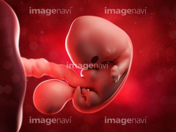
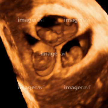
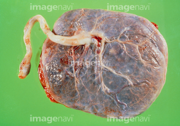
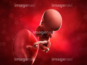
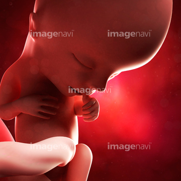
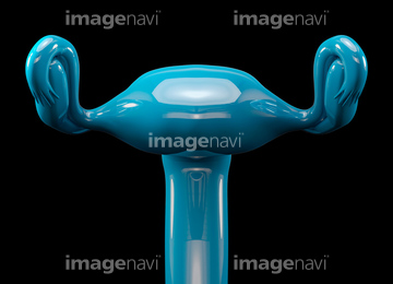
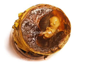
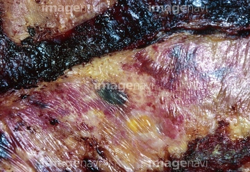
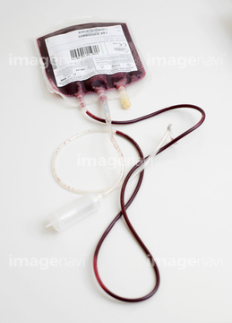
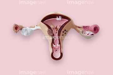
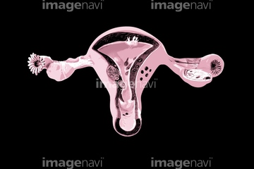
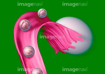
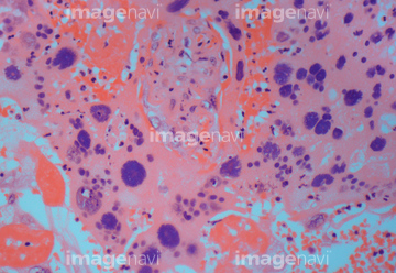
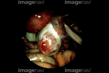
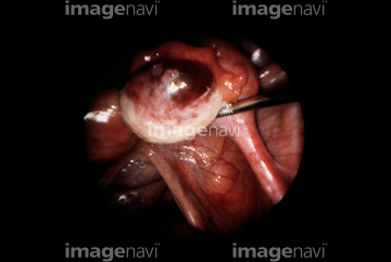
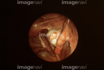
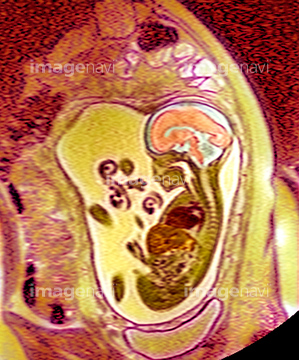
HOME > ژتگ^ > ƒ‰ƒCƒtƒXƒ^ƒCƒ‹ > ‚¨ڈj‚¢ژ–پE’¢ژ– > ”DگPپEڈoژY
10,000Œڈ‚جژتگ^‘fچق‚ھŒںچُ‚³‚ê‚ـ‚µ‚½پB
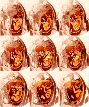
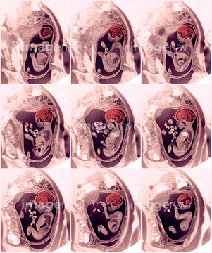
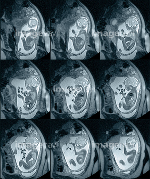
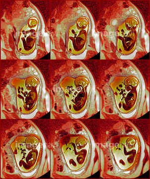
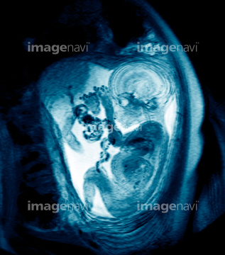
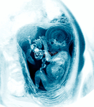
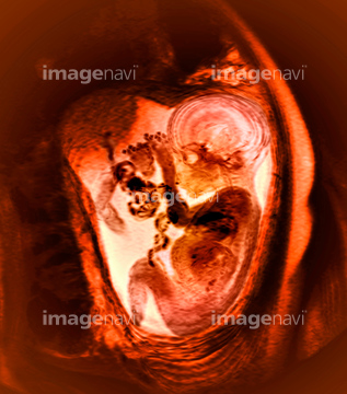
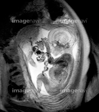
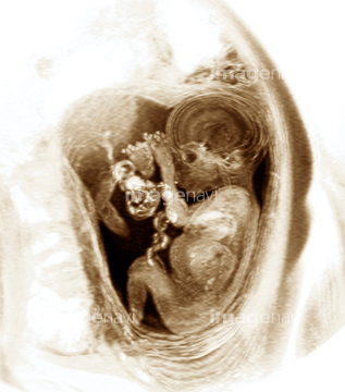
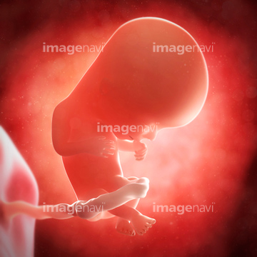
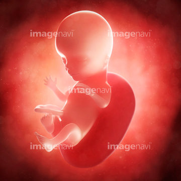
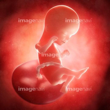
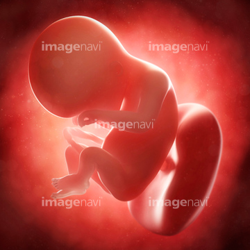
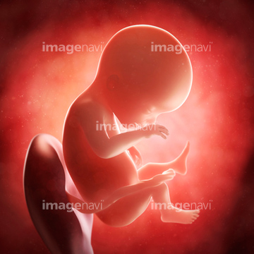
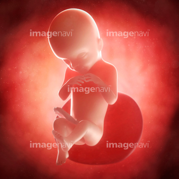
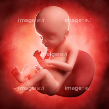
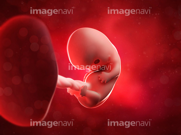
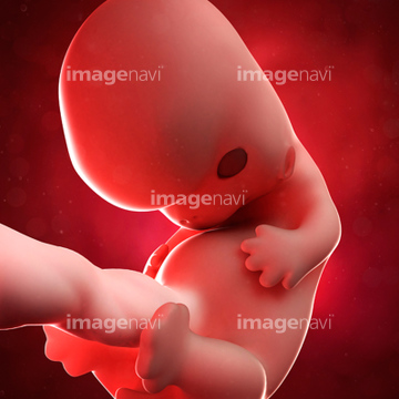
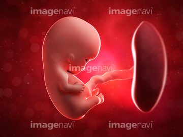
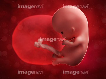
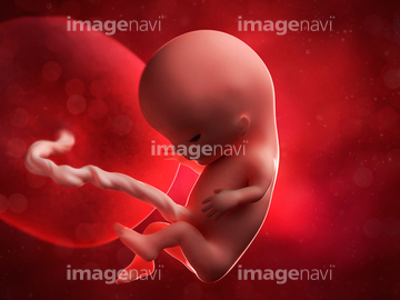
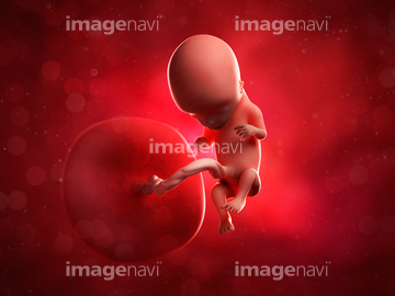
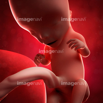
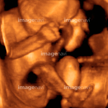
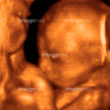
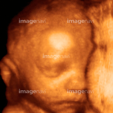
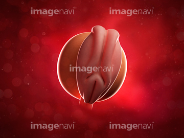
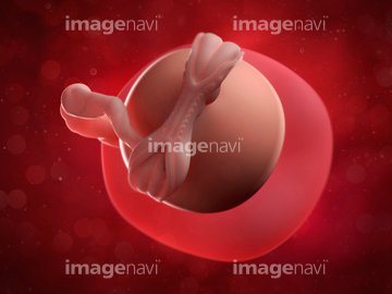
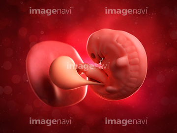
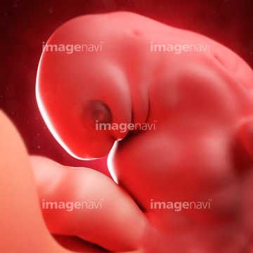
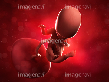
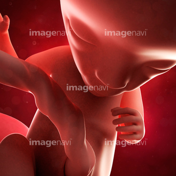
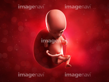
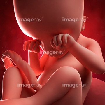
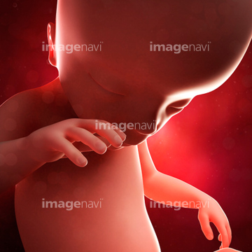
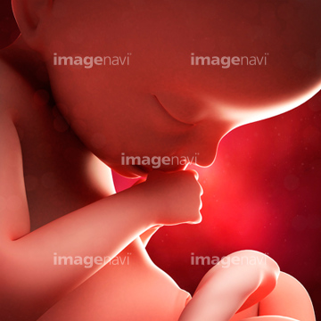
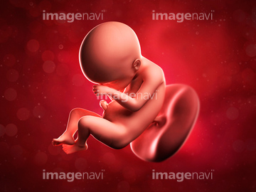
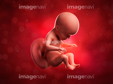
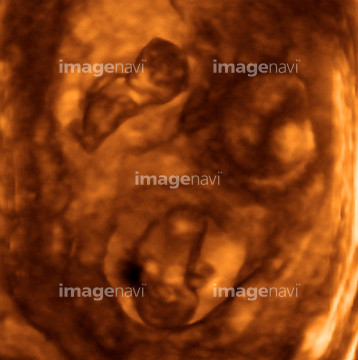
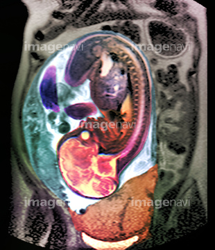
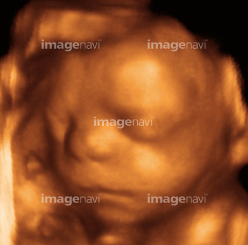
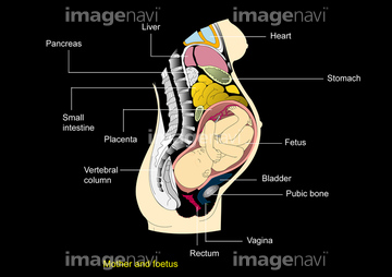
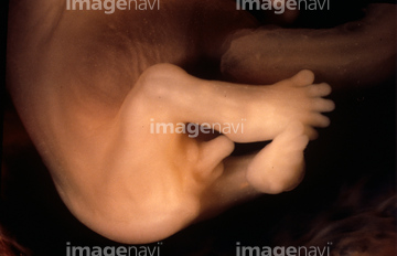
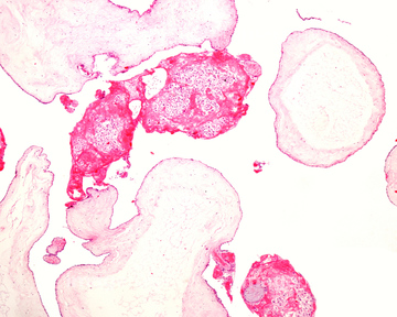
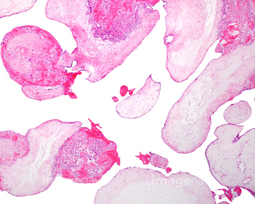
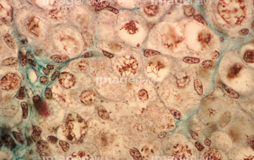
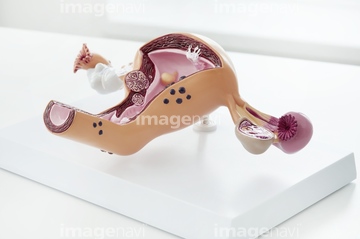
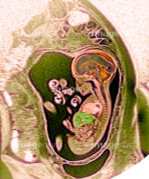
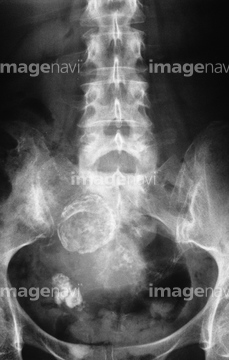
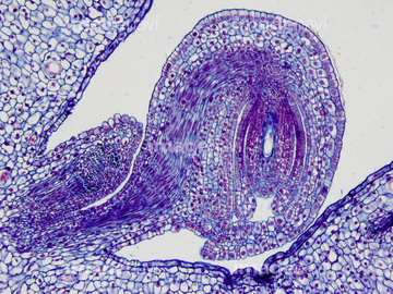
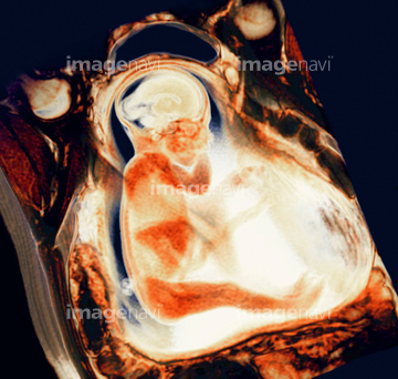
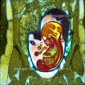
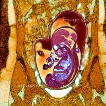
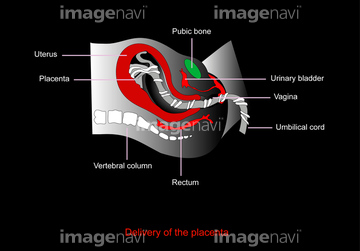
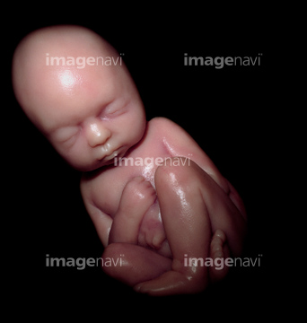
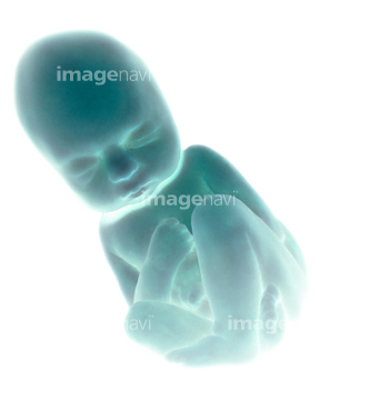
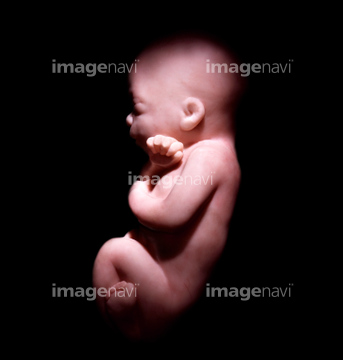
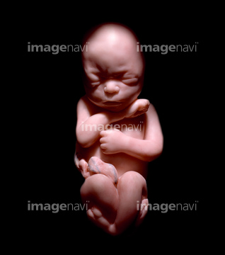
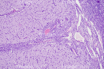
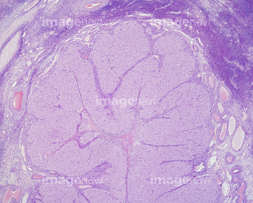
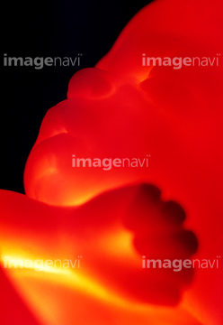
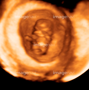
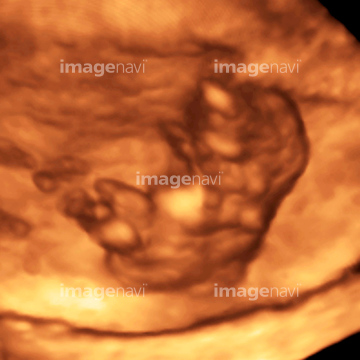
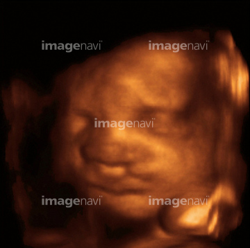
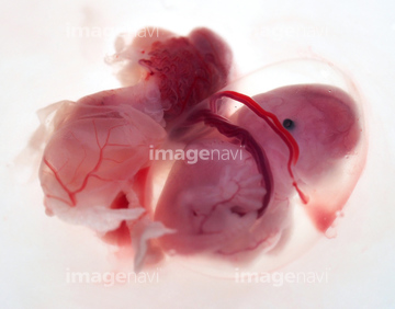
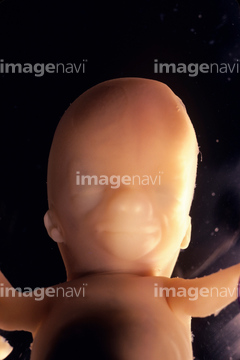
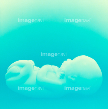
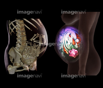
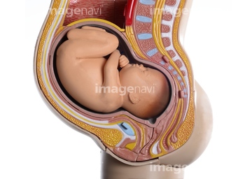
‚±‚جŒںچُŒ‹‰ت‚ة‚حپAFoetus at 18 weeks, artworkپAFoetus at 19 weeks, artworkپAFoetus at 20 weeks, artworkپAFoetus at 21 weeks, artworkپAFoetus at 23 weeks, artworkپAFoetus at 24 weeks, artwork‚ب‚ا‚ھٹـ‚ـ‚ê‚ؤ‚¢‚ـ‚·پB
64021990
64021987
64021988
64021989
64021983
64021984
64021986
64021982
64021985
64063028
64076566
17211353
17211354
17211355

17211356
17211357
17211358
17211359

17211360
17211361
17212692
17212693
17212694

17212695
17212696

17212697
17212698

17212699
17212700
17212701
64022083
64022089
64022095
17212686
17212687
17212688
17212689
17212690

17212691
64008169
64008174
64174342
64021956
64068349
17212702
17212703
17212704
17212705
17212706

17212707
17212708
17212709

17212710
17212711

17212712
17212713

17212714
17212715

17212716

17212717

17212718

17212719
64021951
64021776

64021745

64021777
64084006
64084007
64084016
64084014
64060928

64021746
64021732
64070195

64019426
64084012
64044894
64019429
64019430
64019431

64249536
64249548
64045171
64092975
64013518
64058357
64062787
64019428
64049649
64256471
64022011
64067700
64235064
64022005
64049707
64262599
64262600
64067714
64072741
64072769
64021748
64021749
64021750
64021751
64021752
64021753
64021993
64021994
64062795
64263386
64022127
64021995
64021996
64205878
64205903
64063111
64064569
64006555
64123983
18552255
64084009
64048289
64021991
64021992
64022071
64022072
64224945
64197476
64116646
64116673
64067710
64263169
17256228
17256230
17256252
64234565
64234566
64022076
64008883
64008884
64119230

64157559
64022013
64065617
64062783
64062784
64062785
64062798
64006251
64006281
64006282
64049978
64098225
64072752
64077491
64021938
64062976
64247712
64022078
64090470
64019427
64078933
64171398
| ژںƒyپ[ƒW |
