HOME > 写真 > 科学・テクノロジー > 科学 > DNA・細胞
10,000件の写真素材が検索されました。
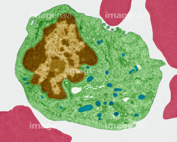
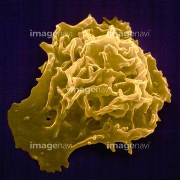
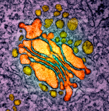
この検索結果には、Photomicrograph of canine eosinphil、Spleen tissue, fluorescence micrograph、Activated plasma cell, TEM、Blood cells, light micrograph、Erythroblast blood cell, light micrograph、Eosinophil white blood cell, TEMなどが含まれています。
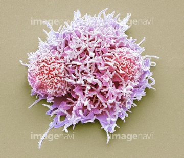
64039289
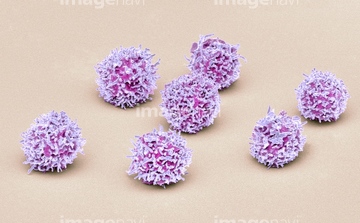
64055762
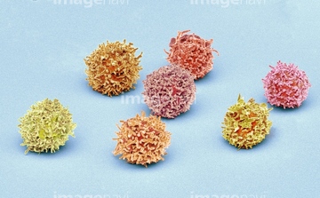
64055775
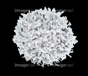
64021403
64196731
64196732
64008096
64008764
64008765
64055761
64062302
64062303
64037572
64037573
64066069
64244243
64055691
64039263
64212972
64212974
64190155
21528573
21528574
64039281
64039282
64039283
64039284
64039285
64084570
64084571
64163462
21528583
17200006
64132607
64132608
64132609
64038830
64038831
64038832
64038834
64038835
64039267
64066067
64163120
64163250
64225179
21528575
21528576
64063021
64063035
64063036
64063037
64063038
64063039
64060811
64060812
64060813
64060829
64060830
64078928
64071063
64071064
64150278
64140892
64140895
64140896
64140898
64132610
64132611
64038833
64221773
64221775
64221776
64221777
17200007
17200058
17200059
17200417
17201388
17201392
17202431
17202432
17207326

17246266
17246269
64011696
64140894
64108903
64113083
64113090
64132310
64132311
64132312
64132317
64132318
64063053
64063054
64063089
64063090
64162633
64162635
64162644
64162649
64162651
64162661
64165074
64165075
64165077
64165080
64165081
64165082
64165083
64165084
64165085
64165086
64165089
64165094
64165095
64165096
64184849
64184850
64184851
64184852
64184853
64205193
64205195
64205382
64205881
64224953
64214806
64214809
64215356
64215357
64215359
64215360
64215956
64217035
64090372
64090464
64090467
64092129
64256627
64256631
64256632
64257061
64257062
64258373

64234412
64234414
64234416
64234417
64234419
64146120
64146125
64146140
64146147
64146149
64145928
64145929
64145956
64145957
64021603
| 次ページ |
