HOME > 写真 > ライフスタイル > 体調 > 健康
10,000件の写真素材が検索されました。
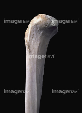
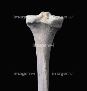
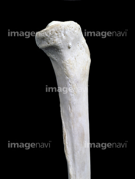
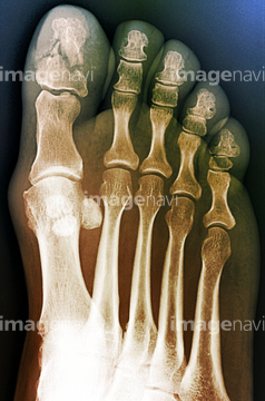
この検索結果には、Tibia, artwork、Proximal view of tibia and fibula、Distal view of tibia and fibula, artwork、Broken ankle, X-ray、Thin woman, MRI scan、Skull in Paget's disease, post-mortemなどが含まれています。
64149490
64149491
64149492
64149493
64149494
64149496
64184381
64264706
64071693
64071695
64071696
64078484
64149495
64071469
64071470
64010078
64078935
64044892
17206849

64083726
64019181
64204483
64220319
64220320
64078139
64078140
20568295
64064634
64064635
64064636
64168725
64264746
64083730
64040050
64070821
64070822
64071951
64071952
64071953
64070829
64070830
64070831
64070832
64070833
64070834
64070835
64070836
64070841
64070842
20559266
20559267
20559268
20559269
20559270
20559271
64045022
64045023
64045024
64044861
16918309
16918340
30342196
30342197
64165250
64187285
64187287
64187288
64084815
64084769
64044871
17218209
64012607
64012608
64012614
64012615
64010085
64243535
64020916
64020917
64020918
64020919
64131735
64109994
17206880

64219640
64220331
64220333
64220334
64168726
64168727
64168728
64045014
64045015
64045016
64045025
64045026

64045027
64194410
64194411
64046179
64075124
64072124
64078371
64078486
64204916
20564383
20566747
20566748
20566749
20566750
20566751
20566752
20566753
20550620
20550621
20550622
20550623
20550624
64044894
64070859
64070860
64045028
64045029
17201776
64021241
64059701
64040391
64040392
51415233
51415234
51415235
51415236
51415237
51415238
51415239
51415240
51415241
51415242
51415243
51415244
51415245
51415246

51415247
51415248
51415249
51415250
51415251
51415252
51415253
51415254
64078963
64078964
64074819
64220322
17249886
17249887
17249888
17249889
17249890
64056052
66000180
64044860
64044870
64084770
64084771
64084772
64084818
64084819
64084820
64044851
64044852
64225355
64225419
64225465
64044891
| 次ページ |
