HOME > 写真 > 花・植物 > 葉
10,000件の写真素材が検索されました。
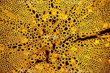
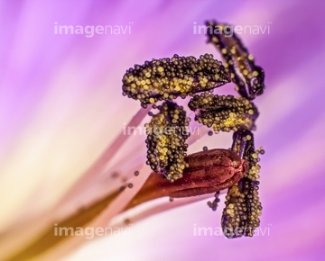
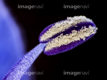
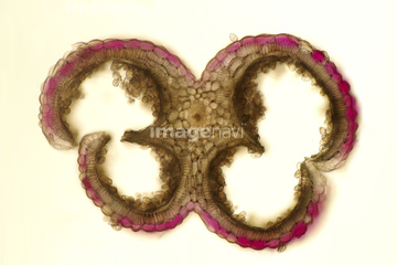
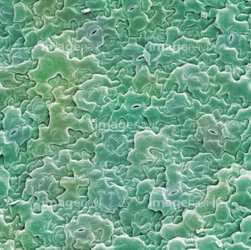
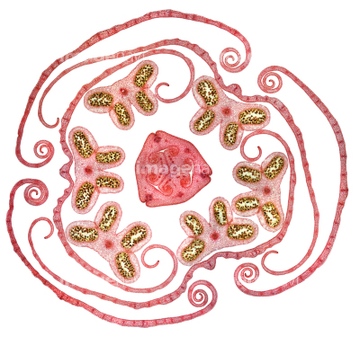
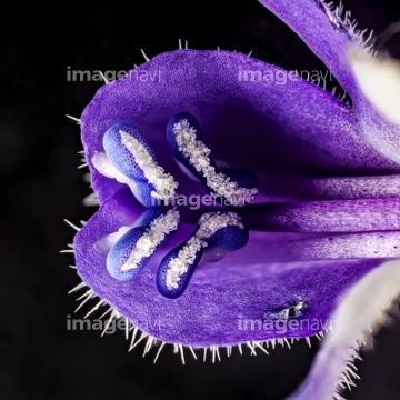
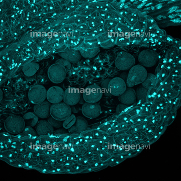
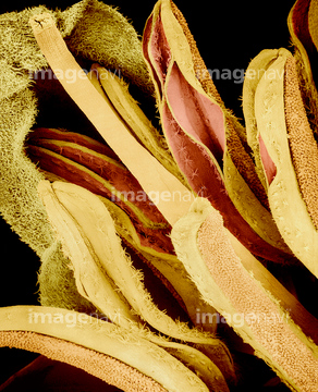
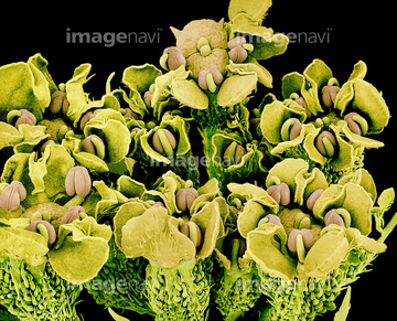
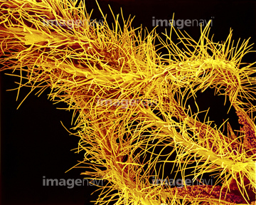
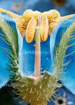
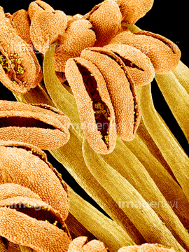
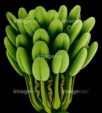
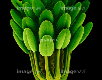
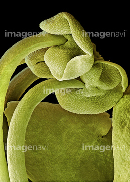
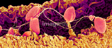
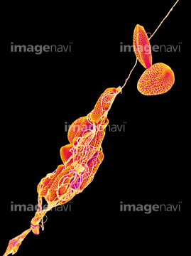
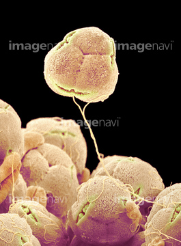
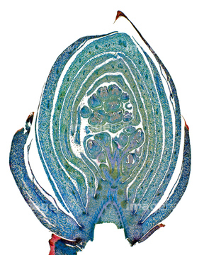
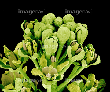
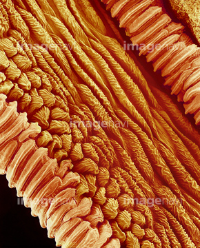
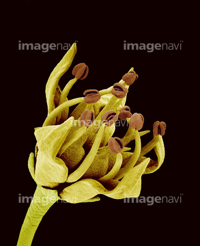
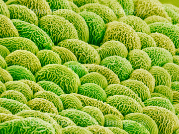
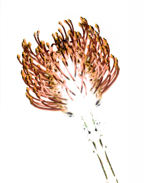
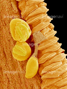
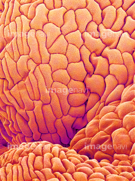
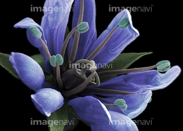
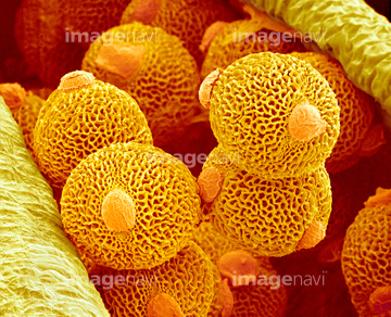
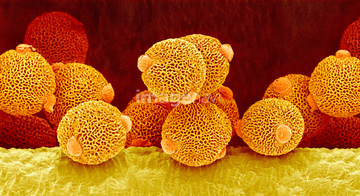
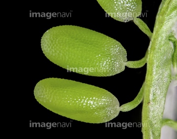
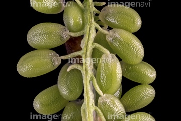
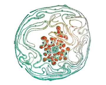
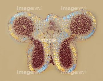
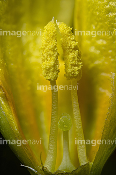
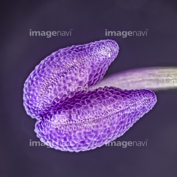
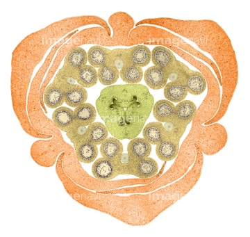
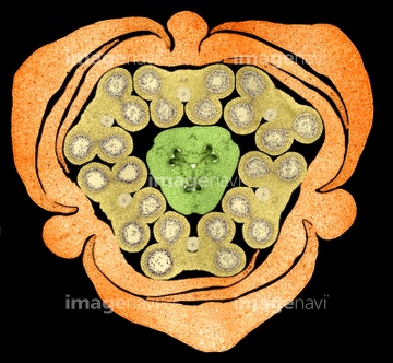
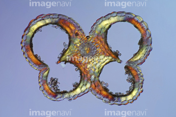
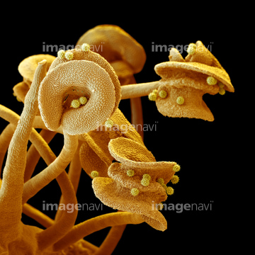
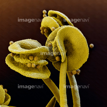
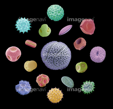
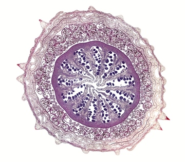
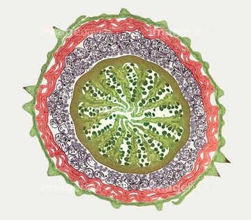
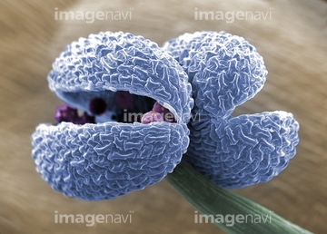
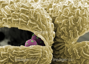
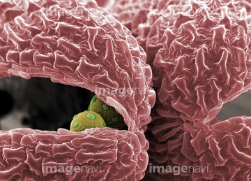
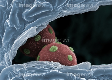
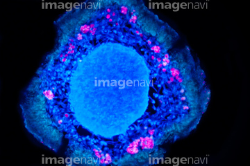
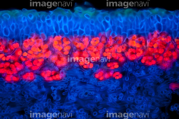
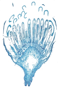
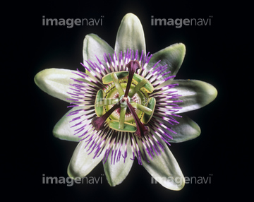
この検索結果には、Columbine flower stamens, SEM、Basil stamens, SEM、Red spider lily pollen, SEM、Azalea pollen, SEM、Lilac flower bud, light micrograph、Stigma of Arabidopsis thalianaなどが含まれています。
64045337
64045340
64045334
64045338
64075078
64075077
64075074
64075075
64045339
64056795
64006244
64056793
64119752
64119753
64119754
64119755
64119756
64006265
64006275
64006510
64006511
64006512
64006270
64171741
64147380
64218399
64064386
64006180
64006246
64006252
64006254
64006255
64006266
64006276
64006280
64006466
64006468
64006469
64006470
64006236
64006249
64006271
64006306
64072454
64072455
64072456
64084023
64056800
64006251
64006250
64006257

64006258

64006259
64006260
64006263
64006289
64006303
64006399
64006179
64006264

64006467
64114731
64008916
64202116
64202117
64208901
64147369
64205190
64006202
64006203
64006304
64006301
64153447
64153448
64153449
64006142
64006253
64006481
64006482
64138238
64138259
64218394
64147371
64147372
64042124
64042126
64045624
64117912
64117913
64117914
64056785
64056786
64056787
64214176
64006310
64006311
64006272
64006290
64006302
64006425
64010987
64153450
64153451
64153452
64153453
64153454
64153455
64153456

64153457
64121541
64121556
64104201
64056789
64056790
64059682
64101247
64101248
64006237
64006309
64006335
64006336
64006346
64006355
64006357
64006372
64061104
64006144
64006181
64006248
64006267
64006268
64006273
64006274
64049249
64073057
64073058
64005977
64006140
64006584
64151007
17206231
17206232
17206233
64202118
| 次ページ |
