HOME > 写真 > 科学・テクノロジー > 科学 > DNA・細胞
10,000件の写真素材が検索されました。
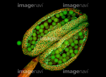
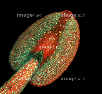
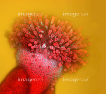
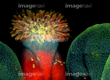
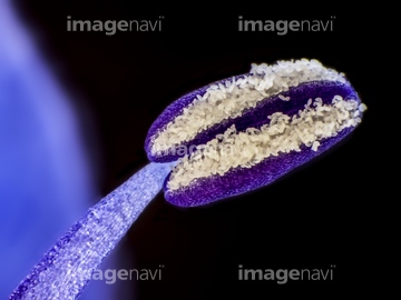
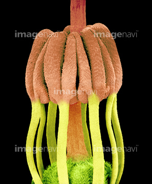
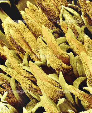
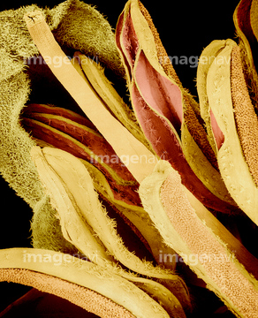
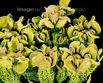
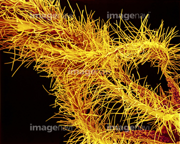
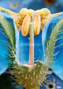
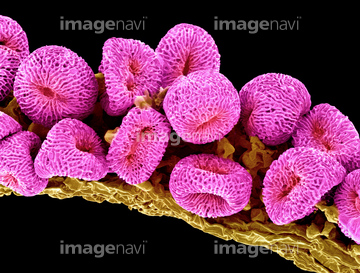
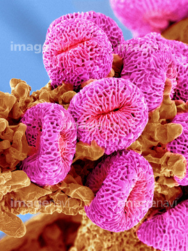
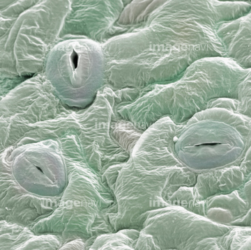
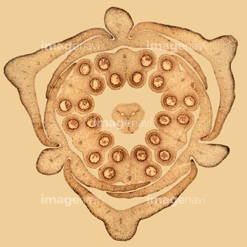
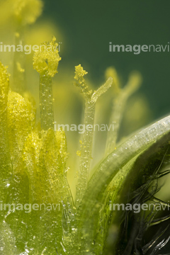
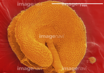
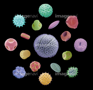
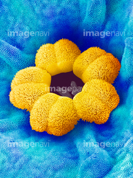
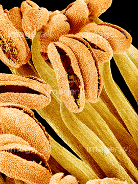
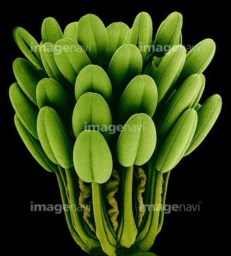
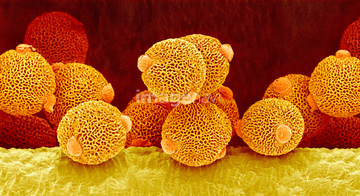
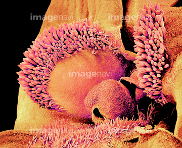
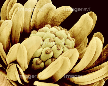
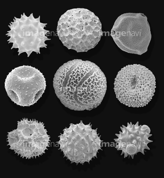
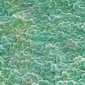
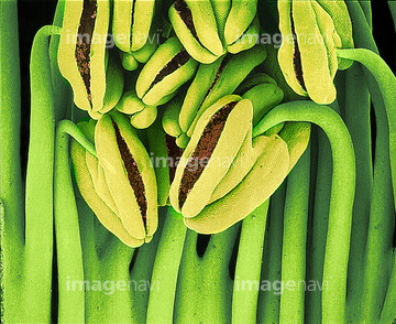
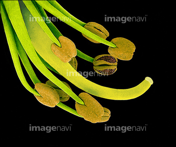
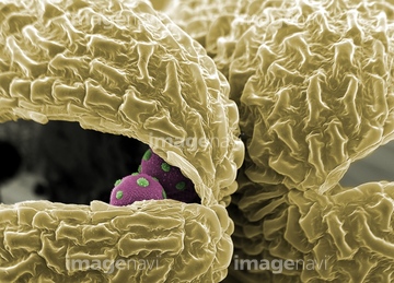
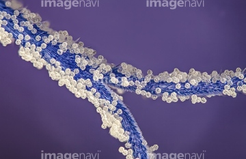
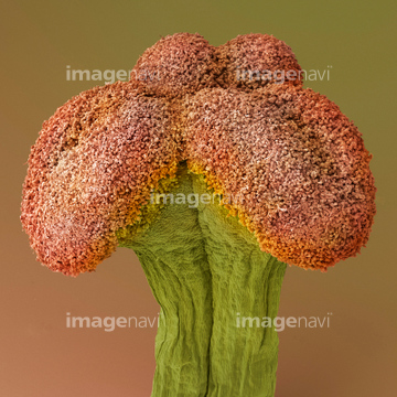
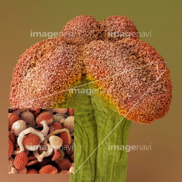
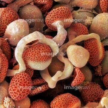
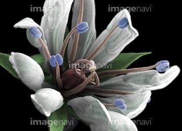
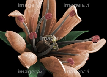
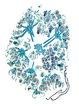
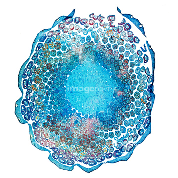
この検索結果には、Open and closed stomates on leaf surface、Lily (Lilium sp.) bud, light micrograph、Daisy flower, light micrograph、Scanning electron micrograph of a hibiscus anther、Pollen grains, SEM、Forget-me-not flower, SEMなどが含まれています。
64045340
64045334
64045337
64045338
64075078
64075077
64075074
64075075
64056795
64006470
64064386
64006246
64006252
64006254
64006255
64006266
64006276
64006280
64006466
64006468
64006469
64056793
64045339
64218399
64006399
64119752
64119753
64119754
64119755
64119756
64006260
64006302
64006304
64153450
64153451
64153456

64153457
64006264

64006467
64205190
64045624
64006142
64006236
64006249
64006253
64006271
64006306
64006481
64006482
64006180
64006244
64006265
64006275
64006510
64006511
64006512
64114731
64155028
64147380
64006270
64006272
64153452
64153453
64153454
64153455
64006289
64006290
64006303
64101247
64101248
64006301
64006425
64006179
64006309
64006335
64006336
64006346
64006355
64006357
64006372
64061104
64147369
64171741
64160944
64006455
64006456
64006479
64218394
64138238
64138259
64006471
64072454
64072455
64072456
64006347
64084023
64092921
64092922
64092923
64089366
64056800
64147371
64147372
20530802
64076435
64076436
64076437
64117912
64117913
64117914
64121541
64121556
64118917
64104201
64213953
64213973
64014623
64014624
64006310
64006311
64006250
64006251
64006257

64006258

64006259
64006263
64010987
64153447
64153448
64153449
64006194
64006204
64006205
64006206
64006237
64077652
64190063
64190064
64190232
64006307

64006338
64008916
64202116
64202117
| 次ページ |
