HOME > 写真 > 人物 > 病気・体調管理
10,000件の写真素材が検索されました。
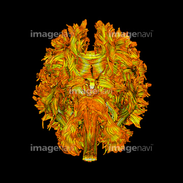
この検索結果には、Memory, conceptual image、Parkinson's disease research、Grey mouse lemur、Studying Parkinson's neurons、Cerebral palsy, DTI scan、Connections between the substancia nigra and the p…などが含まれています。
64061272
64101969
64076175
64076176

64101965
64101966
64101953
64101954
64101956
64101957
64101960
64101961
64101973
64012918
64046001
64101958
64101970
64062611
64062617
64062624
64072631
64072644
17234436
64163099
64101962
20564752
64101967
64186902
64186903

64139123
64101968
64087036
64101955
64101963
64043136
64043137
64043138
64101959
64221143
64221144
53156548
53156549
53156556
53156557
53156558
53156559
53156560
53156561
53156562

53156563
64101971
64101976
64011153
53157256
53157257
53157258
53157259
64065734
64065735
64101977

64043131
64043132
64043133
64043134
64043135
64106797
64106799
64106801
64106802
64106805
64106806
64106807
64106808
64106809
64106810
64093957
64093958
64186898
64186899
64186900
64186901
64216199
64197462
64197463
64048368
17211229
64127301
64127302
64105208
64105210
64220870

64051841
64245985
64257155
64257156
64257157
64216528
64179314
64123110
64101972
64101974
64101975
64043109
64043110
64043111
64043112
64043117
64043118
64043119
64043120
64043121
64043125
64043128
64043129
64043130
64063419
64020504
64129528
17219793
64059439
64059440
64067713
17231560
17231563
17231569
17231576
20530976
20531034
20531035
20536224
17211188
17211191
17211192
17211193
17211194
17211195
20542499
20544119

64043086

64043087
64043088
64043089
64043090
64043097
64043098
64043099
64043100
64043101
64043102
64043103
64043104
64043105
64043106
| 次ページ |
