HOME > 写真 > 科学・テクノロジー > 科学
10,000件の写真素材が検索されました。
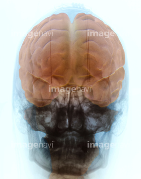
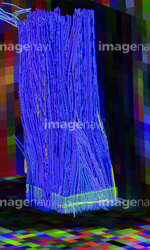
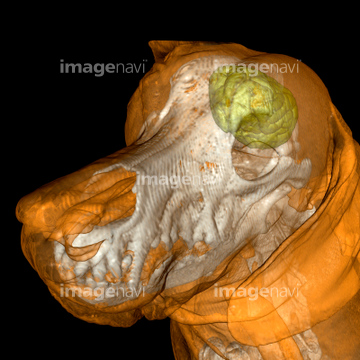
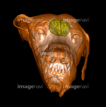
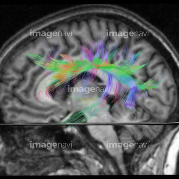
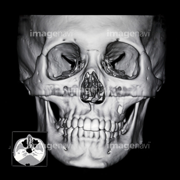
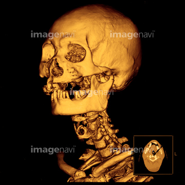
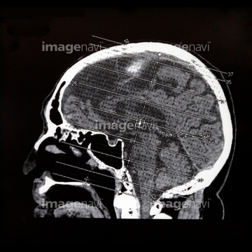
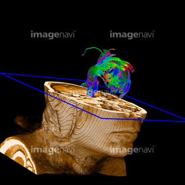
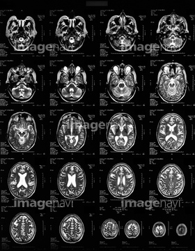
この検索結果には、Fiber tracts of the human spinal cord in a small r…、Dog, 3D MRI scan、Human head, MRI and 3D CT scans、Nine MRI scans of woman's brain、Brain scans, MRI scans、Brain scan, MRI scanなどが含まれています。
64049872
64059701
16918279
16918307
16918314
16918315
16918332
16918335
16918270
64040391
64040392
64021464
64049873
64059785
64059786
16918274
17201401
17201404
64021454
64021468
64021469
64021470
64021471
64021472
64021473
64021478
64021456
64014403
64064693
64064694
64077199
17218191
17218192
17218197
17218200
17218204

17218210
64021447
16918309
16918297
16918278
64035457
64035458
64077202
30064857
64021475
64021477
64021479
20562613
20562614
17247385
17247388
64021451
64021452
64021453
64110133
64043131
16918295
16918304
16918306
64021481
64045014
64045015
64045016
64045025
64045026

64045027
64021297
64021444
64021450
30342251
64021455
64021443
64021445
21508369
16918268
16918284
16918329
64049886
16918273
17218208

64035456
64021405
64066826
64066827
64066833
64066834
64066835
64075025
64075026
64075027
64075028
64043125
64043128
64043129
64043130
51415233
51415234
51415235
51415236
51415237
51415238
51415239
51415240
51415241
51415242
51415243
51415244
51415245
51415246

51415247
51415248
51415249
51415250
51415251
51415252
51415253
51415254
64225127
64019182
64021446
64021448
64021449
10941464
64021304
30342237
64219589
20562609
64021474
64072632
64072640
64072641
64071925
64071926
64071927
64042984
16918323
16918325
64071706
64071707
64063377
64063378
64149487
17201776
20993281

64075285
30304357
30304358
64071942
53156898
53156899
53156900
53156901
64012624
64161164
64161165
64161171
64161172
64161174
64161176
64161177
64161178
| 次ページ |
