HOME > 写真 > 科学・テクノロジー > 科学 > DNA・細胞
10,000件の写真素材が検索されました。
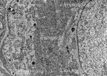
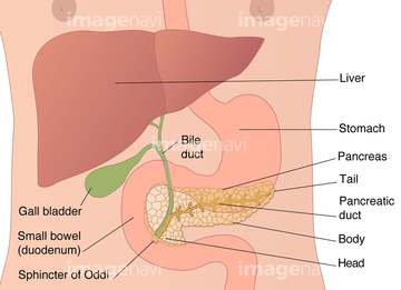
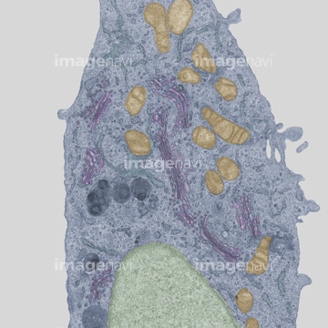
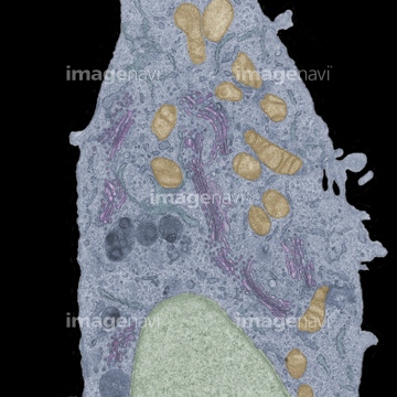
この検索結果には、Pancreatic acinar cell, TEM、Earth's internal structure, artwork、Desmosome cell junction, artwork、Intracellular transport, artwork、Capillary, diagram、Chloroplast structures, artworkなどが含まれています。
64050433
64050434
64048787
64049233
64048789
64050452
64048793
64050439
64048796
64049640
64050446
64060076
64048237
64049234
64049087
64057015
64069867
17212114
64058347
64076254
64076256
64076257
64076266
64076267
64052385
64052386
64050586
64049064
64048261
64049059
17212244
64050437
64048413
64048414
64048833
64049639
64049228
64049229
64049321
64049268
64050447
64247688
64048820
64050253
64052391
64050208
64063632
64063633
64049975
64049060
64057016
64048795
64050440
64071060
64071061
64049317
64056929
64049642
64049648
64145904
64145905

17218196
17218201
64190186
64048786
64060814

64060815
64060816
64208213
64247714
64213924
64076393
64076394
64076398
64076399
64076400
64076401
64048792
64049650
64048286
64048288
64048813
64048818
64049269
64049057
64053254
64048778
64048781
64049327
64150088
64050090
64050158
64050438
64048816
64021602
64050445
64048412
64048415

64049227
64107744
64107746
64021603
64050594
64145894
64145895
64190190
64190191
64190192
64190193
64048788
64048790
64049231
64049232
64051978
64051979
64050453
64145928
64145929
64145956
64145957
64048785
64145898
64145899
64107743
64107745
64051976
64052331
64057018
64050209
64050472
64048259
64048815
64048826
64048827
64052383
64052390
17218189
17218211
64008096
64021596
64021597
64247698
64008572
64021598
64247699
64049272
64049288
64049323
64049324
64049326
64049138
64051977
64049692
64050454
64049646
64049707
64049302
64050442
64048822
64050162
64050206
64048145
64048150
64048256
64048814
64048819
64049067
17229686
17218199
64048780
64048782
64048783
64049706
64049708
64052392
64049287
64050039
64050040
64050448
64145919
64145920
64145896
64145897
64049316
64190896
64190897
64190898
| 次ページ |
