HOME > ژتگ^ > ژY‹ئپEٹآ‹«–â‘è > ƒTپ[ƒrƒX‹ئ > ˆم—أپE•ںژƒ‹ئ
10,000Œڈ‚جژتگ^‘fچق‚ھŒںچُ‚³‚ê‚ـ‚µ‚½پB
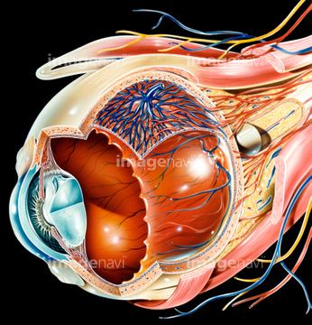
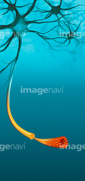
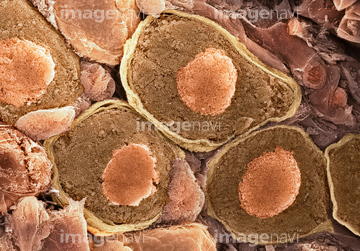
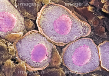
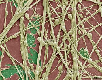
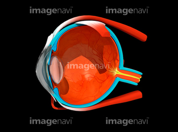
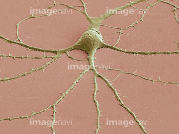
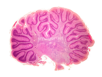
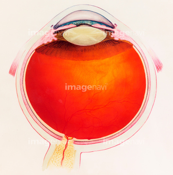
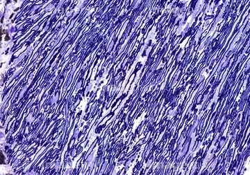
‚±‚جŒںچُŒ‹‰ت‚ة‚حپANerve cells, SEMپASynapse nerve junctions, SEMپAUnmyelinated nerve, TEMپANerve cell, light micrographپAPurkinje nerve cells, light micrographپAEye anatomy‚ب‚ا‚ھٹـ‚ـ‚ê‚ؤ‚¢‚ـ‚·پB
64052131
64052161
64052135
64052157
64052128
64052158
64052136
64052165
64052141
64052154
64052156
64052140
64052134
64052137
64067622
64067623
64086502
64048791
64052133
64067814
64067989
64067618
64067619
64225447
17211187
17211189
17211190
64067813
64042570
64042571
64042572
64048413
64067581
64067582
64067583
17200001
17200002
17200003
64043627
64049961
64039930
64039931
64041144
64041145
64067572
64067573
64068320

64048415

64041146

64059871
64059872
64050697
64050698
64050699
64050700
64067617
64060072
64049990
64048414
64051852
64067713
64012552
64012553
64049075
64067620
64067621
64155006
64059996
64049951
64060220
64043086

64043087
64043088
64043089
64043090
64043097
64043098
64043099
64043100
64043101
64043102
64043103
64043104
64043105
64043106
64043107
64043108
64048781
64049646
64042550
64042556
64042557
30342195
64224994
17231501
17229407
17229557
17229558
64071789
64071790
64148185
64062305
64062306
64067632
64055568
64055569
64256398
64263154
17211185
17211186
17212217
17224400
17224404
17224412
64052145
64063980
64063984
64040604
64040606
64014348

64047399
64049950
64049706
64225303
64060440
17231017
17231560
17231563
17231569
17231576
64106062
64050126
64043091
64043092
64043093
64043094
64043095
64043096
64067637
17201408
17212490
17212491
17212492
17212493
17212494
17212495
17212496
17212497
17212508
17212509
20530976
20531034
20531035
20536224
20542499
20544119
64252594
64225313
64225336
64225342
64225362
64010056

17212516
| ژںƒyپ[ƒW |
