HOME > ژتگ^ > ژY‹ئپEٹآ‹«–â‘è > ƒTپ[ƒrƒX‹ئ > ˆم—أپE•ںژƒ‹ئ
10,000Œڈ‚جژتگ^‘fچق‚ھŒںچُ‚³‚ê‚ـ‚µ‚½پB
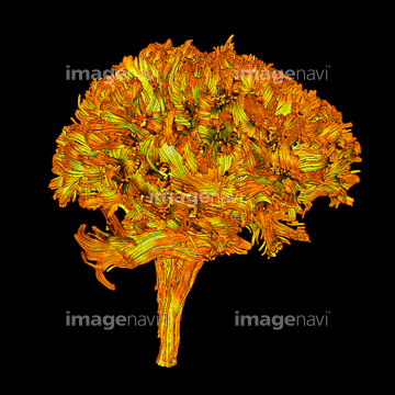
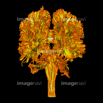
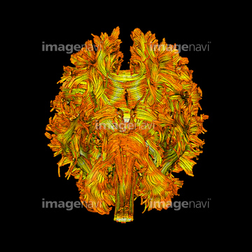
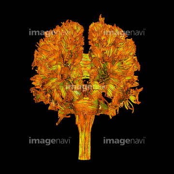
‚±‚جŒںچُŒ‹‰ت‚ة‚حپAWhite matter fibres, DTI scanپAMyelinated nerve, TEMپAInner ear structures, 3D MRI scanپASagittal view of the brain and spinal cord on a T1پcپANerve fibres, SEMپANerve cells, computer artwork‚ب‚ا‚ھٹـ‚ـ‚ê‚ؤ‚¢‚ـ‚·پB
64052158
64052156
64052161
64052157
64052131
64052136
64052165
64052135
64052141
64052154
64052128
64052137
64052140
64052134
64048783
64048790
64049984
64052109
64067989
64048781
64086502

64052133
64048791
64048413
64050697
64050698
64050699
64050700
64049075

64033011

64043086

64043087
64043088
64043089
64043090
64043097
64043098
64043099
64043100
64043101
64043102
64043103
64043104
64043105
64043106
64043107
64043108
64067581
64067582
64067583
64153321
64168010
64168011
64168012
64192565
17200001
17200002
17200003
64043627
64049961
64049990
64048414
64051852
64049646
64039930
64039931
64041144
64041145
64041146
64042550
64042556
64042557
64067572
64067573
64067622
64067623
64067713
64068320
64048415
64148185
30342195
64055568
64055569
20531049
20531050
20531051
64010535
64040391
64040392
64052145
64040604
64040606
64049950
64049951
64042570
64042571
64042572
64067620
64067621
64159962
17231017
17231560
17231563
17231569
17231576
64059216
64043091
64043092
64043093
64043094
64043095
64043096
64050126
64060220
64067618
64067619
64202541
64202542
64010056
64060440
17201417
64112209
64043131
64257155
64257156
64257157
64257167
64086310

64049591
64072632
64072640
64072641
64049706
64039926
64039927
64039928
17212490
17212491
17212492
17212493
17212494
17212495
17212496
17212497
17212508
17212509
64050720


64050731
17212516
17231501
17229407
17229557
17229558
17229566
17229567
17229568

64047399
64050571

64019977
64067813
64067814
17211185
17211186
17211187
| ژںƒyپ[ƒW |
