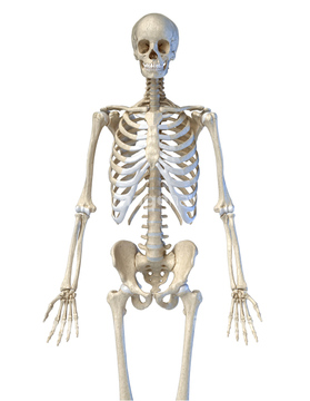
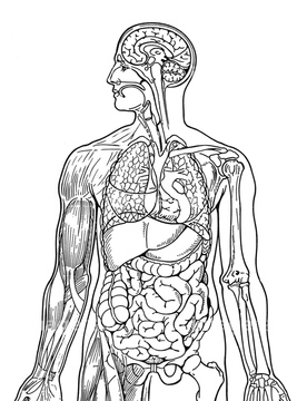
HOME > 写真 > イラスト・CG > 人物
10,000件の写真素材が検索されました。
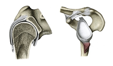
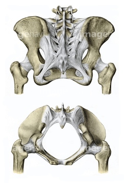
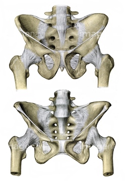
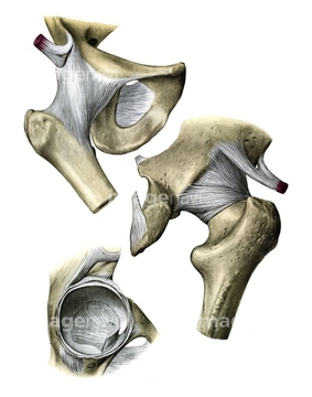
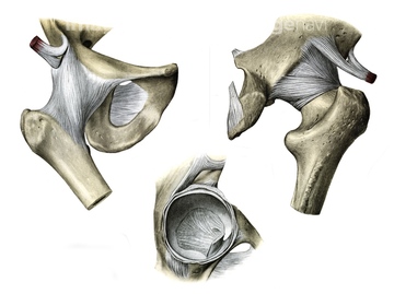
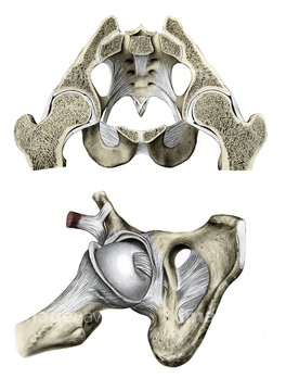
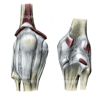
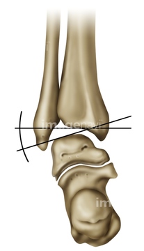
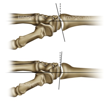
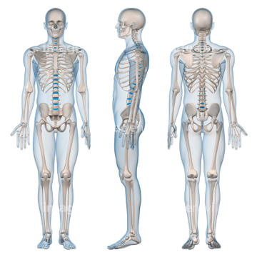
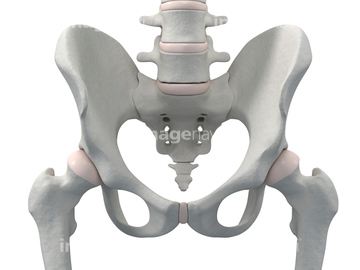
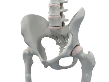
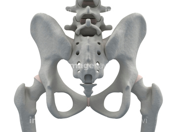
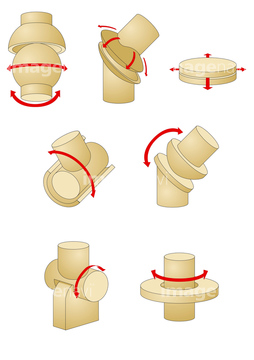
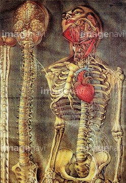
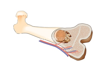
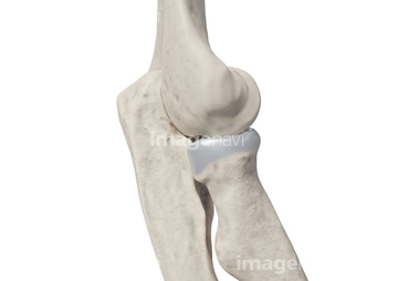
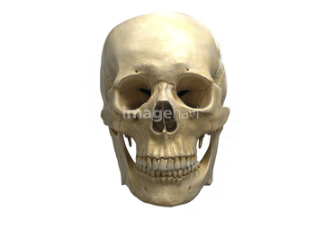
この検索結果には、Pelvis with ligaments, illustration、Hip joint, illustration、Pelvis and hip joint, illustration、Knee joint, illustration、Ankle joint movement, illustration、Wrist joint movements, illustrationなどが含まれています。
64055415
64046466
64048024
64064220
64046462
64089132
64084430
64084431
64084432
64044528
64046505
64064233
64044454
64047324
17202039
17202041
64047911
17207575
17207651
64064910
64124647
64183425
64183436
64183443
64183444

64183445
64183448
64183449
64183450
64183451
64183452
64183453
64183454
64183455
64183456
64183457
64183459
64183460
64183461
64183464
64183466
64183467
64183468
64183469
64183470
64179241
64179242

64179243
64179295
64217404
64183411
64085407
17201824
17201825
17206886
17206887
17206888
17207576
17207577
17207578
17207579
17207580
17207581
17207582
17207583
17207646
17207648
17207649
17207650
17207652
17207653
17207654
17207655
17207656
17207657
17207658
17207690
17207691
17207692
17207882
17207883
17207884
17207885
17207886

17207887
17207888
17207889
17207910
17207911
17207912
17207913
17207914
17207915
17207916
17207917
17207918
17207919
17207920
17207921
17207922
17208011
17208012
17208033
17208120
17208121
17208122
17208123
17202040
17202042
17202051
17226637
17226645
17226669
17226681
17226682
17226742
17226315
17226549
20506341
20506343
20506346
20506377
20506379
20506392
20506395
20506402
20506408
20506427
20506438
20506444
20506448
64045427
64045433
64046500
64046501
64046502

64046503
64046504
64075468
64160454
64160455
64187247
64187248
64187249
64109798
64194379
17240154
64177910

64110015
64110042
64044532
64096252
64049439
64049441
40313583
64088159
17227374
64120781
64120782
64232075
64217385
64217780
| 次ページ |
