HOME > 写真 > 科学・テクノロジー > 科学 > DNA・細胞
10,000件の写真素材が検索されました。
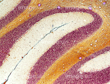
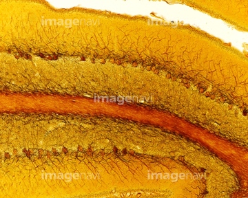
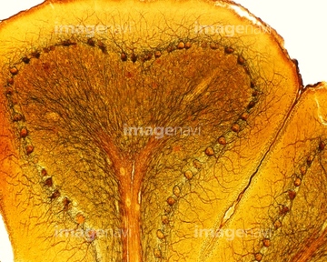
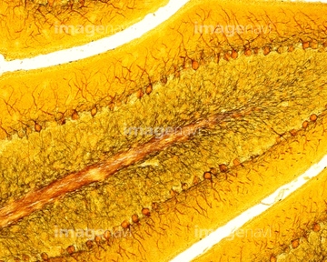
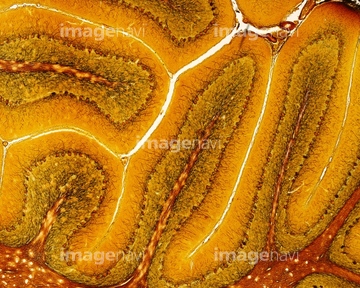
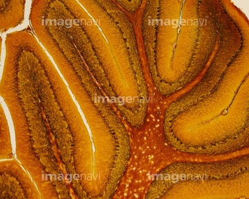
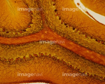
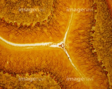
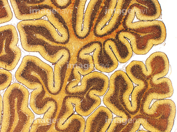
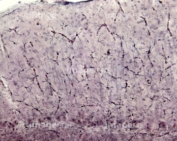
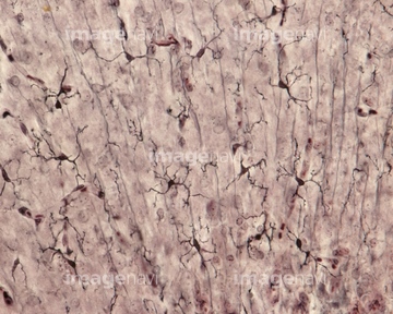
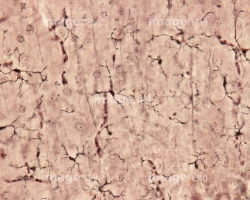
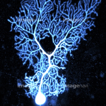
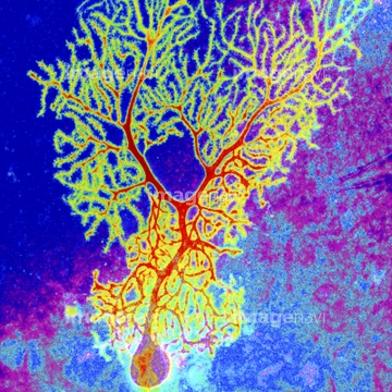
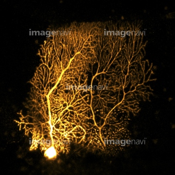
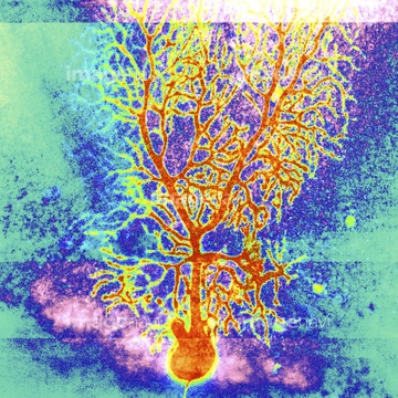
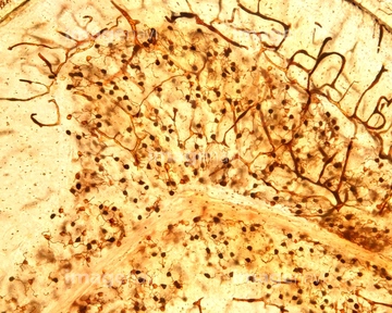
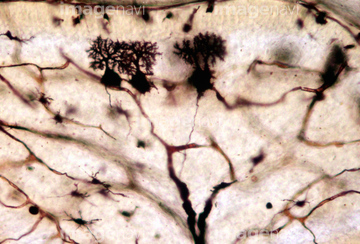
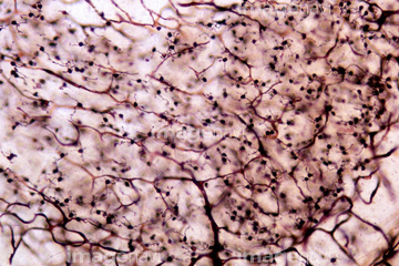
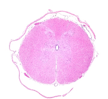
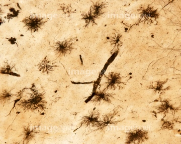
この検索結果には、Cerebellum tissue, light micrograph、Cerebellum, light micrograph、Microglia, light micrograph、Cerebellum, light micrographs、Cerebellar cortex, light micrograph、Purkinje nerve cell, confocal micrographなどが含まれています。
64055569
64055568
64055570
17211188
17211191
17211192
17211193
17211194
17211195
64194276
64194277
64194278
64194279
64194280
64194281
64194282
64194283
64194284
64194285
64194286
64194287

64194289
64199165
64199166
64199167
64199168
64213886
64213887
64213888
64260249
64234732
64234733
64043091
64043092
64043093
64043094
64043095
64043096
64194270
64194288
64197464
64234719
64234720
64234721
64234722
64234724
64234725
64234731
64223998
64223999
64013293
64060930
64250027
64251231
64194271
64194272
64194273
64194338
64197465
64093965
64093982
64194290

64260024
64180131
64180132

64206758
64206759
64248269
64043086

64043087
64043088
64043089
64043090
64043097
64043098
64043099
64043100
64043101
64043102
64043103
64043104
64043105
64043106
64043107
64043108
64048791
17231560
17231563
17231569
17231576
64159970
17219793
53157050
64194267
64194268
64194269
64194311
64197467
64197468
64197470
64197471
64197469
64159971
64174847
64055606
20542525
64090460
20531049
20531050
20531051
64077341
64077342
64077343
64257120
64194261

64194303
64194339
64194340
64194341
64197466
64060440
64258370
64258371
64254566
| 次ページ |
