HOME > 写真 > 人物 > ビジネス > 学者・研究者
10,000件の写真素材が検索されました。
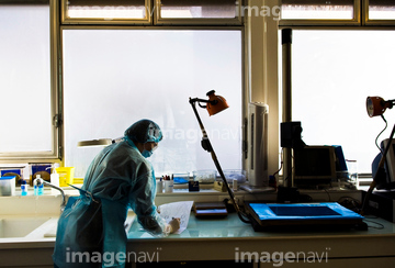
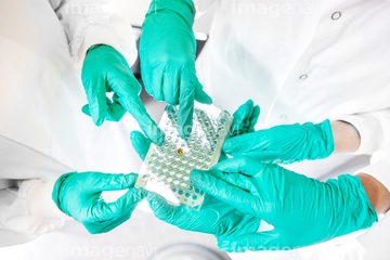
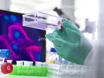
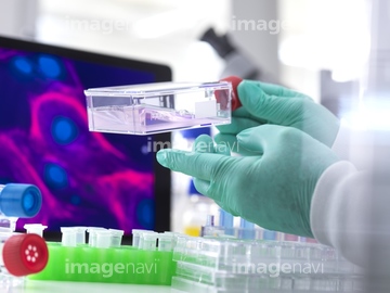
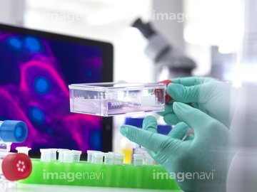
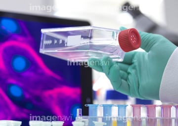
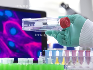
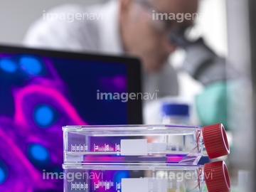
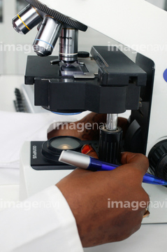
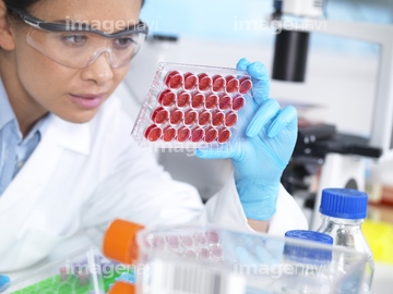
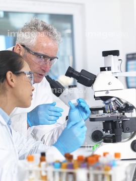
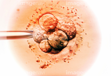
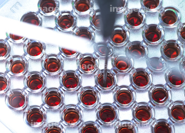
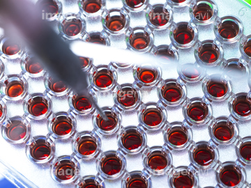
この検索結果には、Kidney tissue, confocal micrograph、Squamous epithelium, confocal micrograph、Squamous epithelium、Spectrophotometer analysis、Stem cell research、Cell researchなどが含まれています。
64045905
17251109
17251110
64044047
64222900
17239621
17239622
17239623
17239624
17239625
17246298
17251082
17251083
17251084
17251085
64125374
17284926
17284927
17284928
17284929
64059673
64059674
20500082
20500113
20510205
20510207
20510227
20570181
20570182
64096365
64105557
64074135
64085919
64050042
17246313
17246314
64059670
64059680
64125390
17280155
17280156
17280157
17280158
17280173
64125375
64125376
20510200
20510201
20510202
20510203
20510204
20510206
64223193
64071257
64071304

64118378
64118493
64086354
20536005
20536009
20536010
64011645
17246286
64125388
64188513
64188514
64144127
64125391

30445780
64223102
64223103
64086379
53136137
40010791
40010793
40010796
40010797
17272433
17280174
17280747
17251100
17251101
17251102

64071180
64071186
64071244
64085920
17251093
17251094
17251095
17251096
20500073
20500074
64058066
64048771
20518658
20518659
20518685
20518686
20518687
20536019
20536022
20537648
64140649
64140655
20500065
20500066
64187413
64188464
64221792
20500076
20500087
20500088
20500089
64046001
17251113
17251114
20536006
20536007
20536008
17260856
64059676
17251115
64015191
21565176
21565177
21565178
20500106
17232475
20582770
20582776
20582777
20582778
20582779
40010795
64172494
64172495
64172496
64161164
64161165
64161171
64161172
64161174
64161176
64161177
64161178
64161179
64161181
64161182
64161183
64161187
64161194
64161200
64161201
64161204
64050041
| 次ページ |
