HOME > 写真 > 医療・福祉 > 医療 > 医療器具
10,000件の写真素材が検索されました。
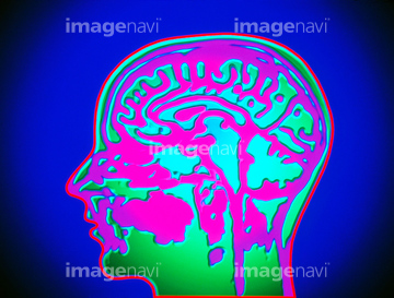
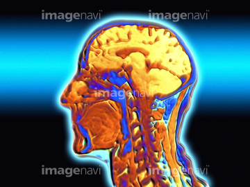
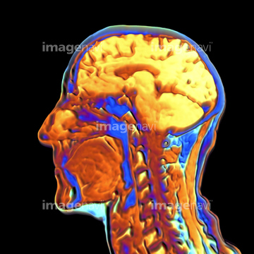
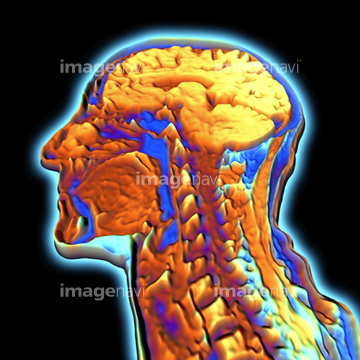
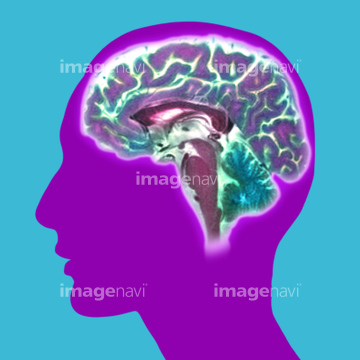
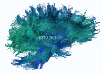
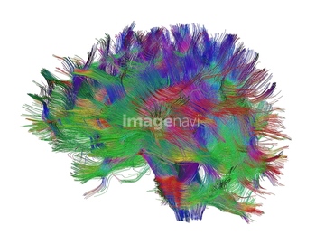
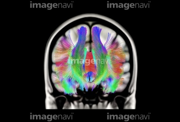
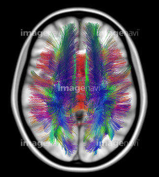
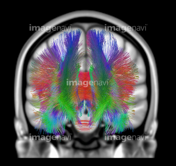
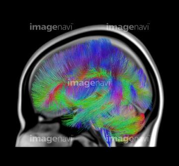
この検索結果には、False-colour MRI scan of the head, axial section、Brain limbic system, 3-D MRI scan、Ventricles of brain, MRI、Brain, 3-D MRI scan、Brain scan, MRI scan、MRI scan of human brain, close-upなどが含まれています。
64059701
64049872
64040391
64040392
64071942
64059785
64059786
17206962
17206963
64063731
64063749
16918270
64064693
17201401
64071941
64204278
64204279
64204280
64204281
64204282
17201404
64064694
17218191
17218192
17218197
17218200
17218204

17218210
64021473
64045028
64045029
64078080
64078081
64225127
64021451
64021452
64021453
64021468
64021469
64021470
64021471
64021472
64049873
64014403
64021447
64021454
64021456
64021478
16918314
16918315
16918332
16918335
64149562
21508369
30064857
17200425

51415233
51415234
51415235
51415236
51415237
51415238
51415239
51415240
51415241
51415242
51415243
51415244
51415245
51415246

51415247
51415248
51415249
51415250
51415251
51415252
51415253
51415254
64225000
16918297
64166783
16918279
16918307
30342251
64072632
64072640
64072641
17218208
64021475
64021477
64021479
64071706
64071707
64071925
64071927
16918273
64021481
64063377
64063378
64021443
64021444
64021445
64021446
64021448
64021449
64021450
64021455
64219589
64084816
64071926
64042984
64043131
64046178
64046179
17223829
17223837
17223840

64071447
64159954
64149559
64149565
64149566
64203166
64159953
64049886
64066826
64066827
64066833
64066834
64066835
64045019

64045020
64045021
64077199
16918336
20566620

64090770
64021466
64021467
64021464
64021474
64263862
64013523
64161164
64161165
64161171
64161172
64161174
64161176
64161177
64161178
64161182
64161187
64161200
64161201
64161204
64172494
64172495
64172496
64159967
64059784
30304357
30304358
64043160
20582138
| 次ページ |
