HOME > 写真 > 医療・福祉 > 医療 > 医薬品
10,000件の写真素材が検索されました。
この検索結果には、CT scan 84 year old male with Alzheimer's disease.…、Toolbox.、Grey mouse lemur、Antibody Alzheiimer's treatment、Post-menopausal uterus, X-ray、Diseased brainなどが含まれています。
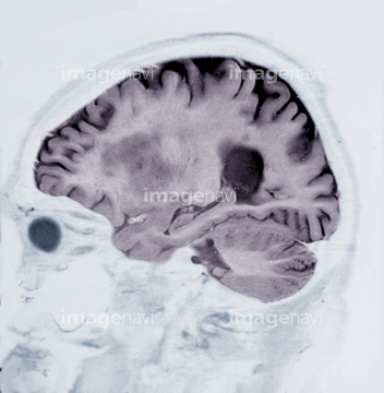
64062617
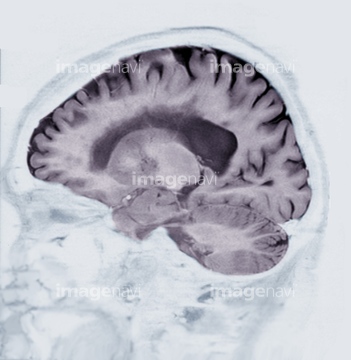
64062611
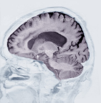
64062624
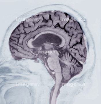
64072631
64072644
64163408
53157256
53157257
53157258
53157259
64186902
64186903
64210309
64210310
64210311
53156548
53156549
53156556
53156557
53156558
53156559
53156560
53156561
53156562

53156563
64012918
64020504
64186898
20510183
20510184
64106797
64106799
64106801
64106802
64106805
64106806
64106807
64106808
64106809
64106810
64042984
17264976
17264977
64186899

53157029
64087036
17237936
64105212
64021754
64093542
20564681
64139114
64139120
64139121
64124609
64139123
64179314
64216528
64093957
64093958
64018484
64165330
64165331
64165332
64165333
64165334
64165335
64165336
64165337
64165338
64165339
64165340
64165341
64165342
64165343
64165344
20510185
20510186
20510187
20510188
20510189
20560090
64008418
64127301
64127302
64186900
64186901
64165637
64055054
17261256
17239188
17239189
64124610
64123113
64165345
64165346
64165347
64165348
64165349
64165472
64165473
53156979

64065916
64127299
17237929
64194355
64194356
64194357
64194358
64101973

64125399
64139124
64139125
64139126
64250735
64250736
64216199

17208414
64018481
64076175
64076176
64061272
64019879
64123112
64263769
64157116

64008416
64008417
64008419
64008420
64165487
64112235
64186259

64188268
64188269
53156978
20582428
20568330
17291210

64165630

64126395
64123111
| 次ページ |
