HOME > 写真 > 医療・福祉 > 医療 > 病院・クリニック
10,000件の写真素材が検索されました。
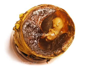
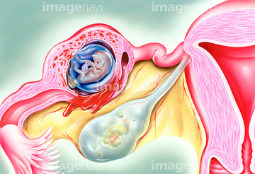
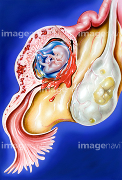
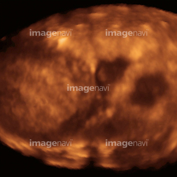
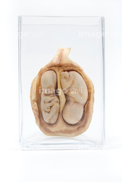
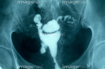
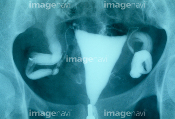
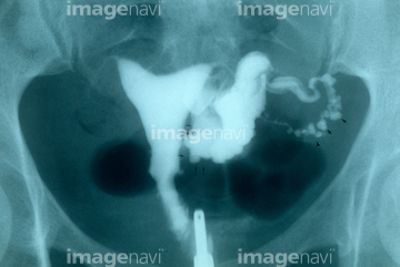
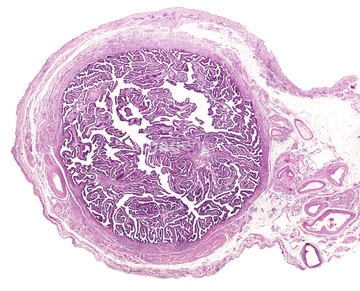
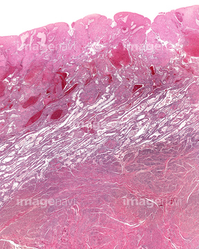
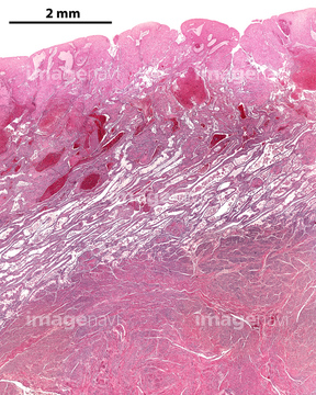
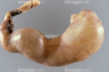
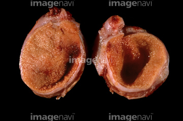
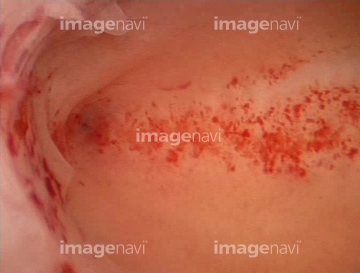
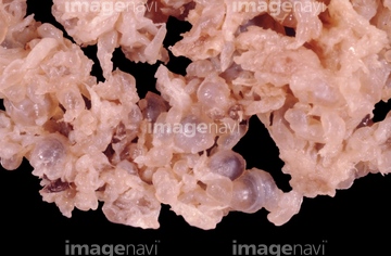
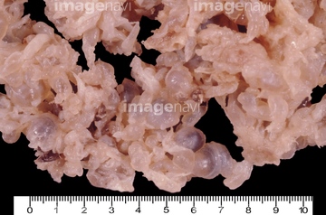
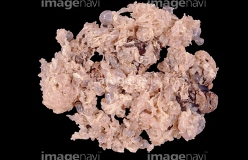
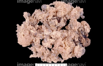
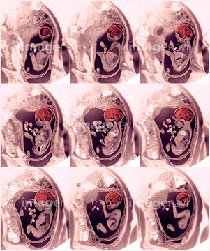
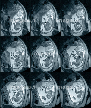
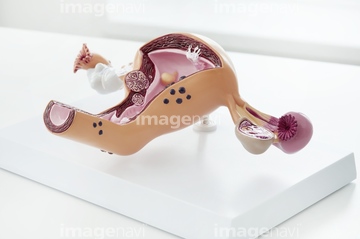
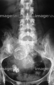
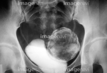
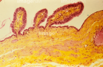
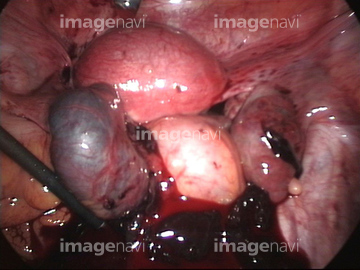
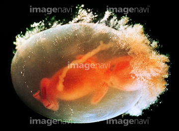
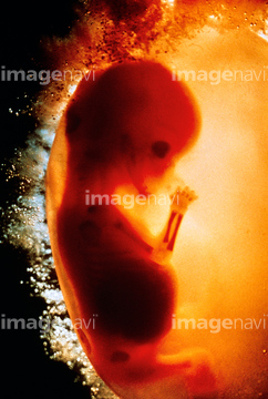
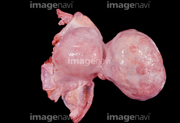
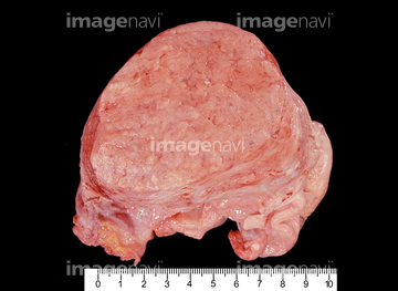
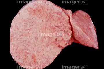
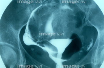
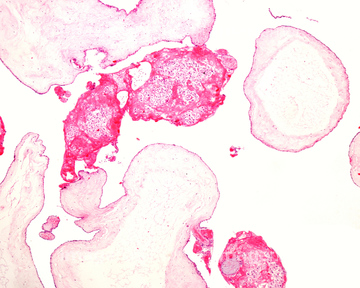
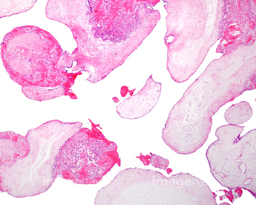
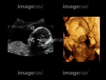
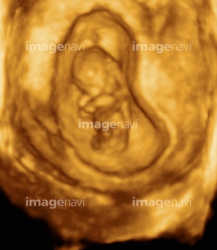
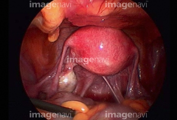
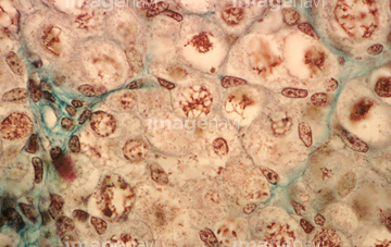
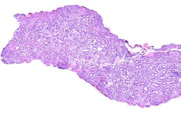
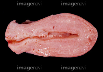
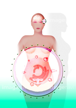
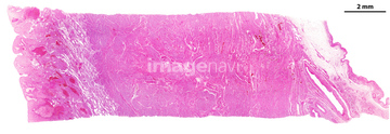
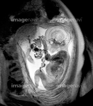
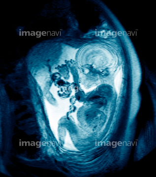
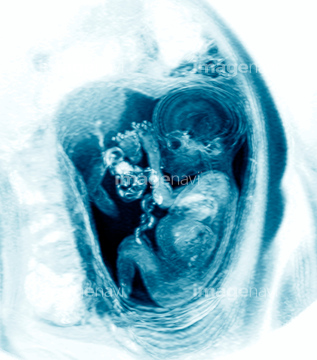
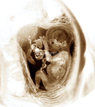
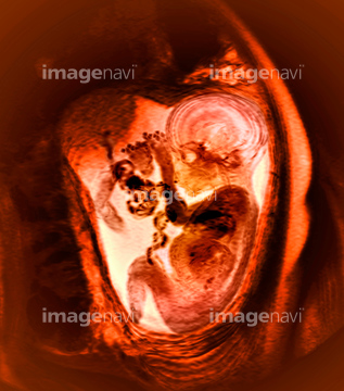
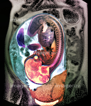
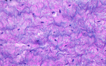
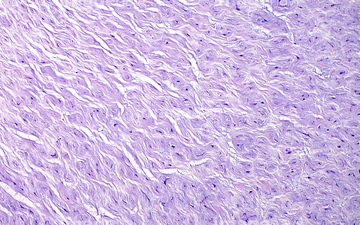
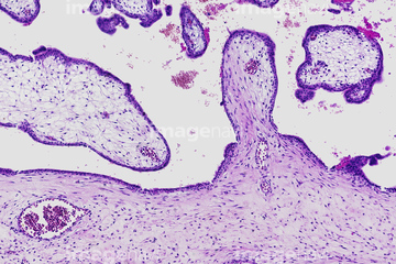
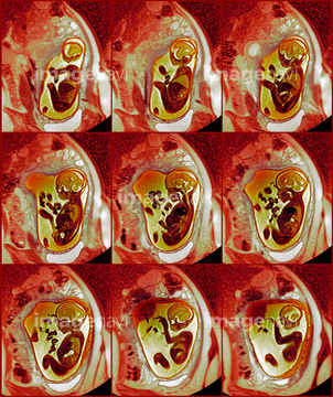
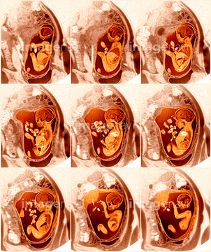
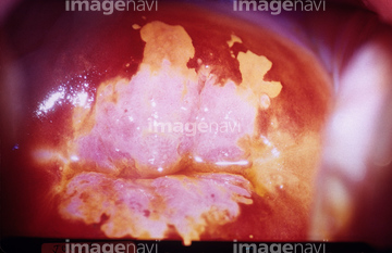
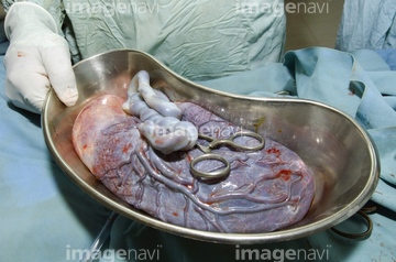
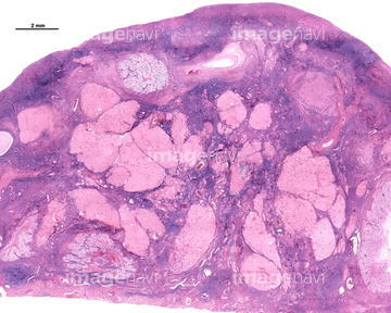
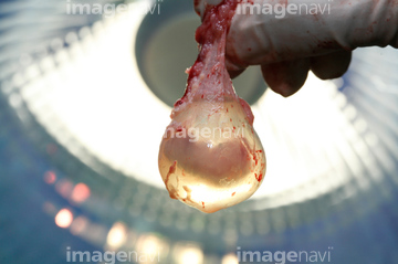
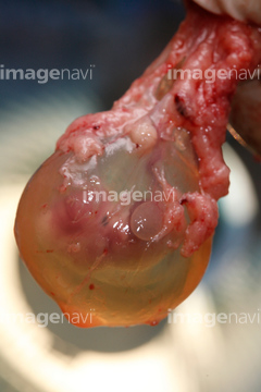
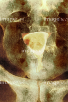
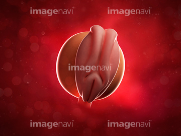
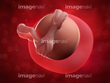
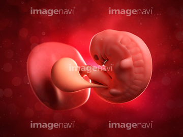
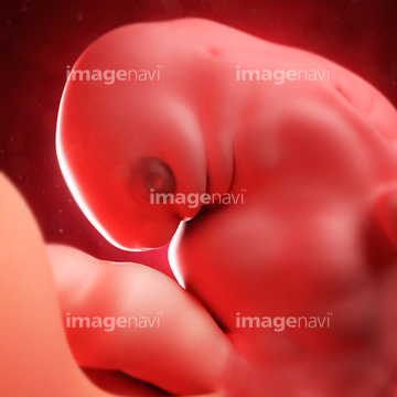
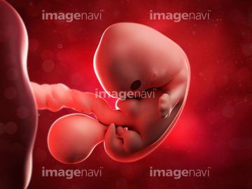
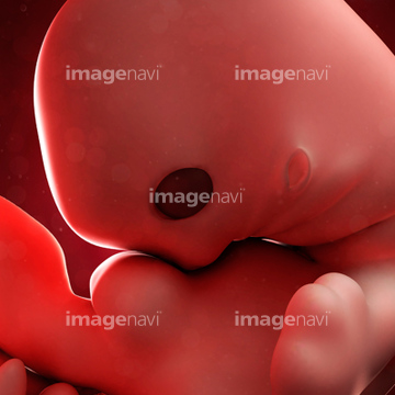
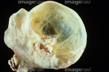
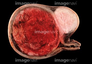
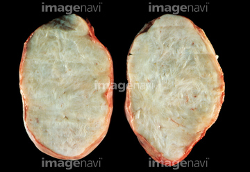
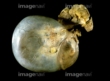
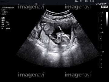
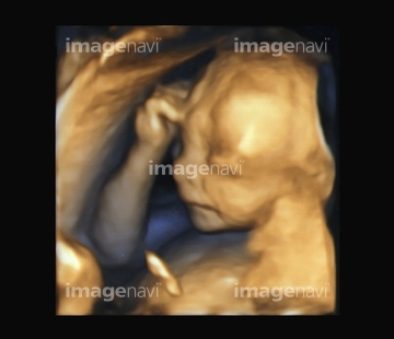
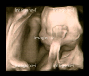
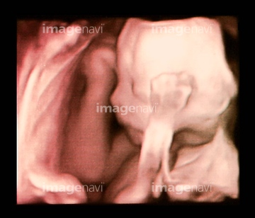
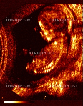
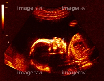
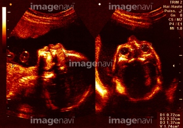
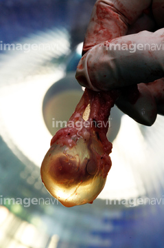
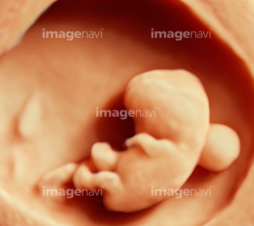
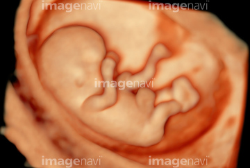
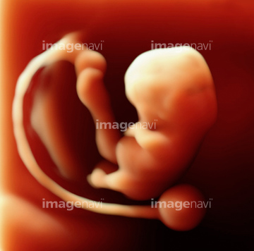
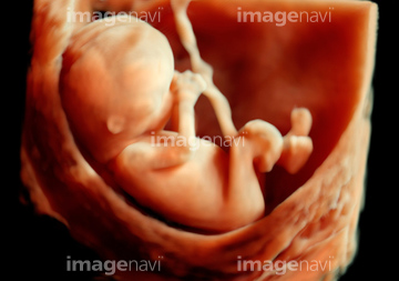
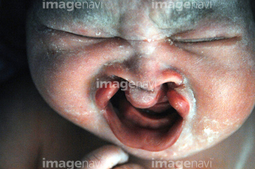
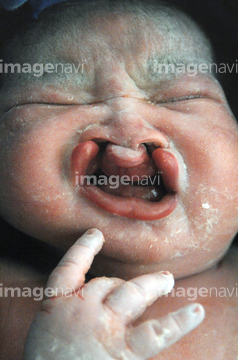
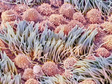
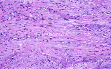
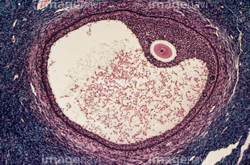
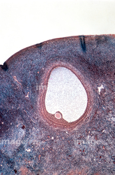
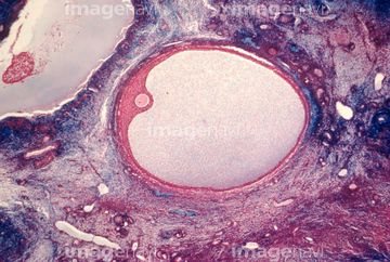
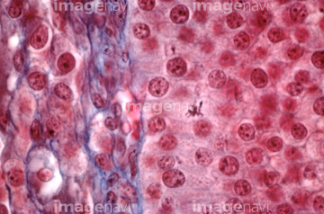
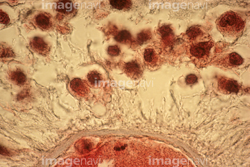
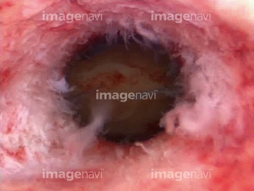
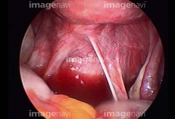
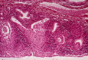
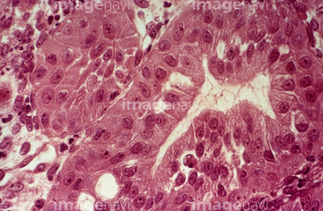
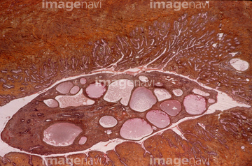
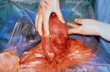
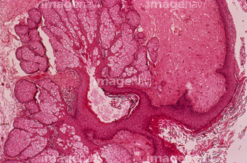
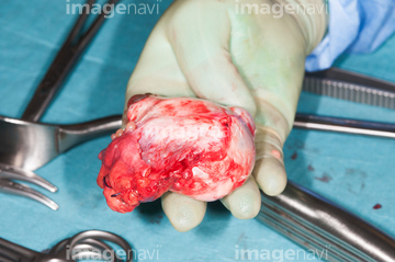
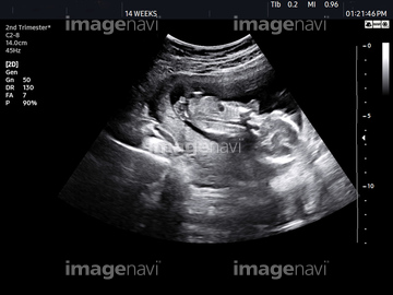
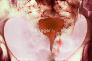
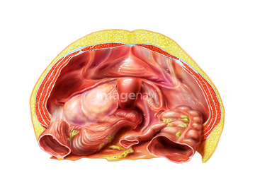
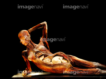
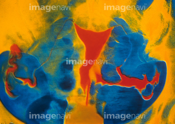
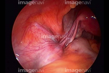
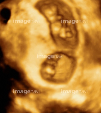
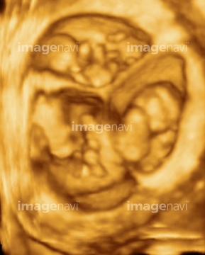
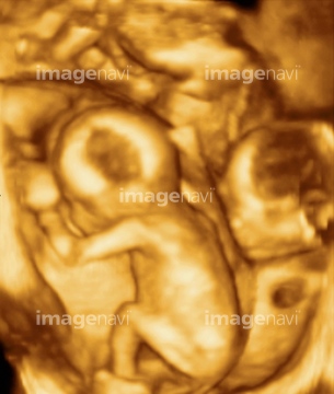
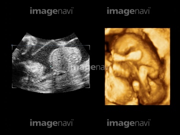
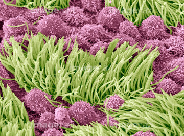
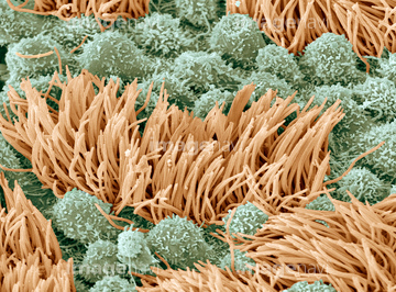
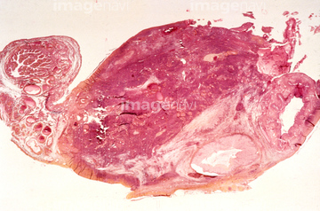
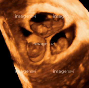
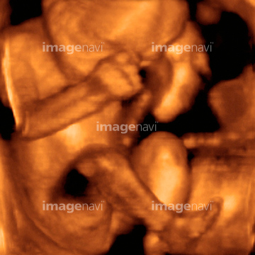
この検索結果には、Placenta, light micrograph、Foetus, MRI scans、Corpus albicans, light micrograph、Cervical erosion、Genital wart, endoscope view、Dermoid ovarian cystなどが含まれています。
64062787
64067985
64067986
64013521
64045171
64062788
64062791
64065614
64065775
64219696
64219697
64219698
64219699
64021987
64021988
64062795
64062796
64065621
64013524
64063369
64063370
64257169
64257170
64257171

64062789
64262599
64262600
64154889
64257172
64067710
64021982
64021983
64021984
64021985
64021986
64044894
64256471
64235064
64021989
64021990
64065601
64065583
64065774
64065983
64065987
64222358
64172525
64222253
64222254
64059784
17212686
17212687
17212688
17212689
17212690
17212691
17212692
17212693
17212694
17212695
17212696
17212697
17212698
17212699
17212700
17212701
17212702
17212703
17212704
17212705
17212706
17212707
17212708
17212709
17212712
17212713
17212714
17212715
17212716
17212717
17212718
17212719
17212710
17212711
17211353
17211354
17211355

17211356
17211357
17211358
17211359
17211360
17211361
64022011
64222234
64222235
64222236
64222237
64222238
53122746
64065591
64065618
64065619
64065623
64065624
64065784
64065984
64191818
20564293
64264220
64189962
64189963
64189964
64189965
64189966
64222231
64064578
64064580
64064581
64064582
64064583
64064584
64064585
64064586
64064587
64260587
64243640
64222661
64222662
64056751
64263141
64063350
17201435
64174342
64063028
64062937
64062938
64062969
64062970
64062971
64062972
64062973
64062974
64062975
64062976
64062977
64062982
64154881
64154883
64065589
64065615
64065782
64065872
64065873
64264222
64059783
64060063
64065576
64154885
64154886
64154887
64154888
17202455
17202456
64065594
64021956
64022083
64022089
64022095
| 次ページ |
