HOME > 写真 > イラスト・CG > CG > 3DCG
10,000件の写真素材が検索されました。
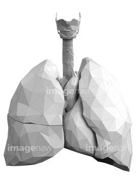
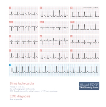
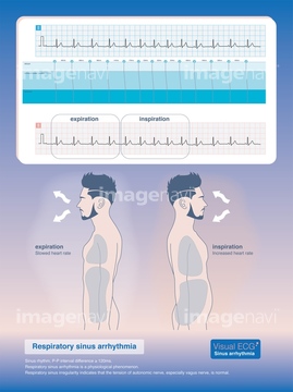
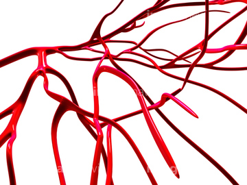
この検索結果には、Fatty heart, illustration、Heart, abstract illustration、Heart and lungs, abstract illustration、Sinus tachycardia, illustration、Respiratory sinus arrhythmia, illustration、Arteries, computer artworkなどが含まれています。
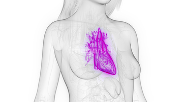
64064233
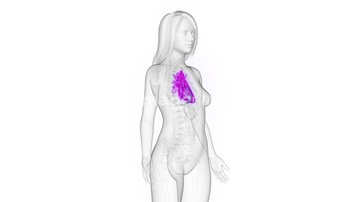
64245359
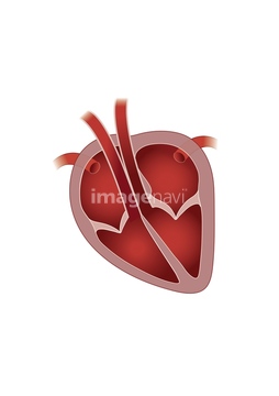
64062349
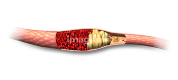
64055873
20549888
20549895
20551254
20551255
20551256
20551257
20551258
20551259
20551260
20551261
20551262
20551263
20551264
20551265
20551266
20551267
20551268
20551269
20551270
20551271
20551272
20551273
20551274
20551275
20551276
20551277
64065176
17207697
17207708
17207718
17207723
17207734
64047876
64044454
64047324
17273434
17273454
20551281
64222133
17202037
64062953
17207947
17207961
17207969
17289326
20551250

20551251
20552832
20552833
20552834
20552835
20552836
20552838
20552840
20552841
20552842

20552851
20552852
20552853
20552854
20552855
20552856
64061192
64061193
64061200
64061201
17285353
17285354
40530037
40530039
64044968
17273442
64047833

64040283
64217158
17273436
64245354
64064220
64166144
64047493
17202034
17202035
17202036
64074930
20552427
20552440
20552451
20552466
20552475
20552526
20552527
20552555
20552556
20552557
20549813
20549814
20549815
20549816
20549817
20549818
20549819
64076493
64047920
64047494
64222122
64222139
64222140
64222142
64222153
64252437
64252438
64163452
64146591
64122619
64219098

17207953
17207992
17208023
64122827
64122828

64122830
64122833
64122838
64063127
64064094
64047360
29148513
17200083
17200085
17200137
17200138
17208245
17208257
64145369
64145370
64145384
20576896
20576899
64241925
64065132
20579366
20576277
20576282
16929666

20551018
20551025
20551028
| 次ページ |
