HOME > 写真 > 科学・テクノロジー > 科学 > DNA・細胞
10,000件の写真素材が検索されました。
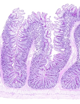
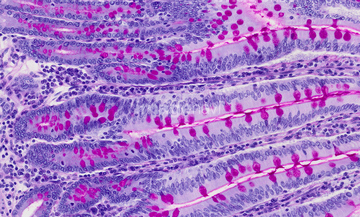
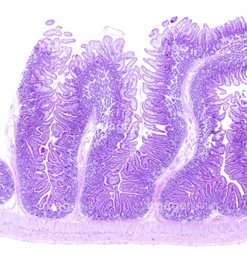
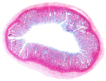
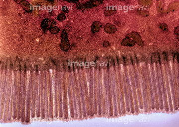
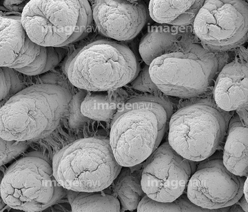
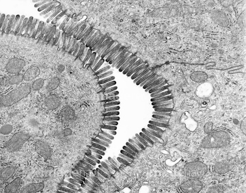
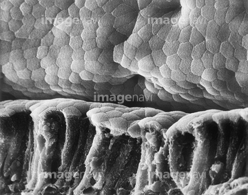
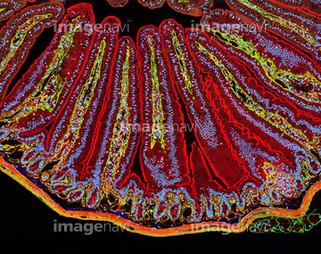
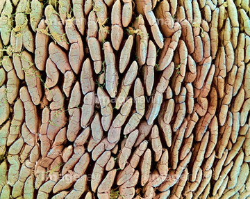
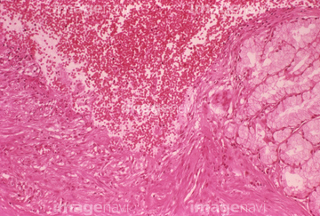
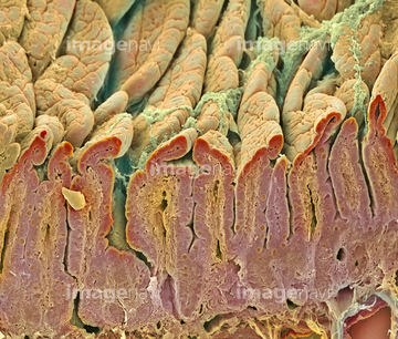
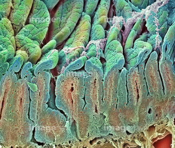
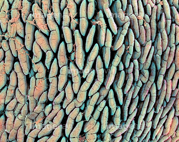
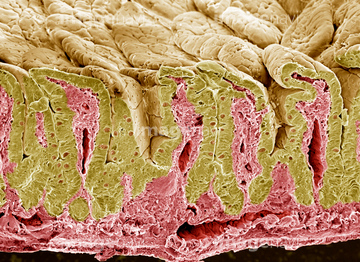
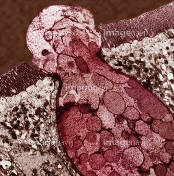
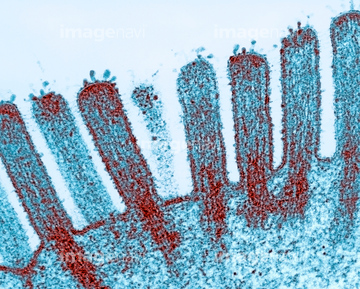
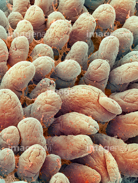
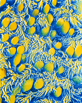
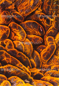
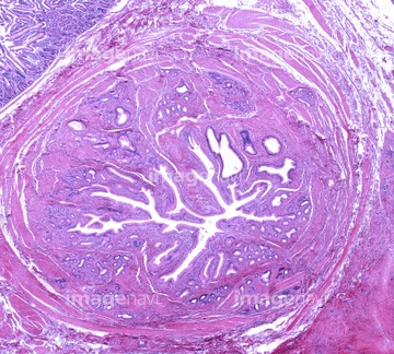
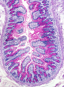
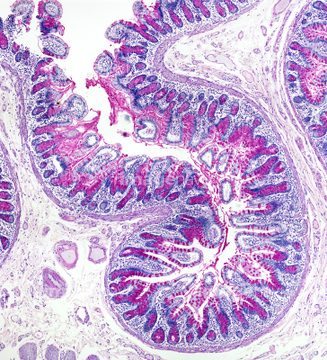
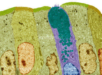
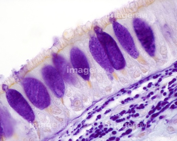
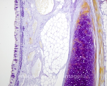
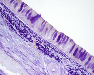
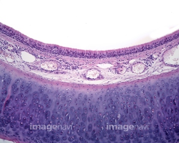
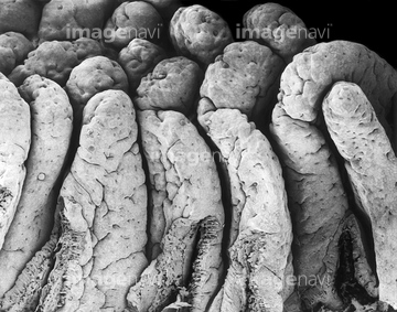
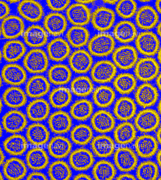
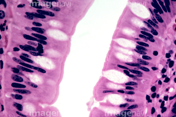
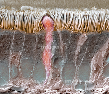
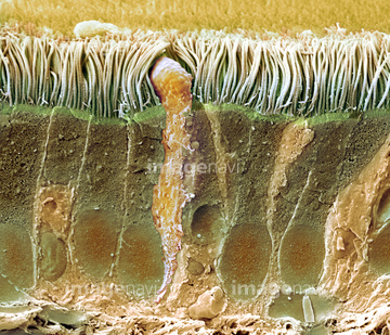
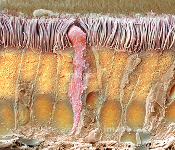
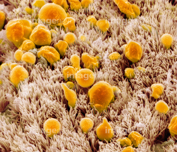
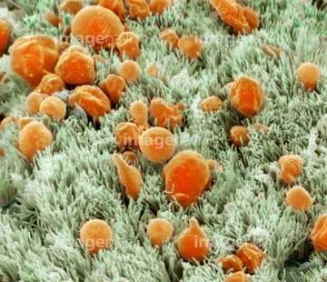
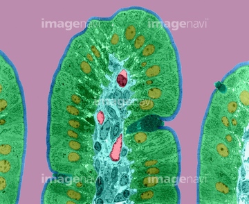
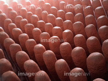
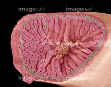
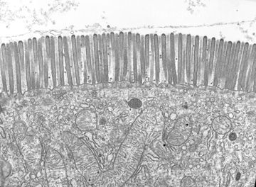
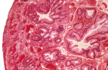
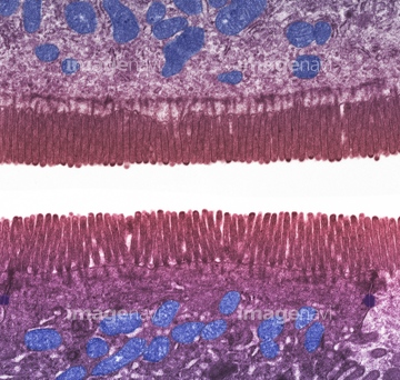
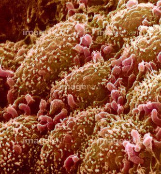
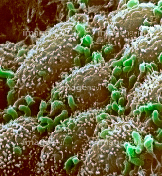
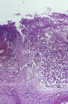
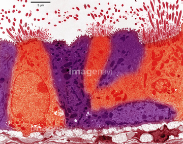
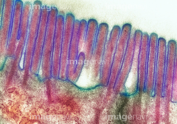
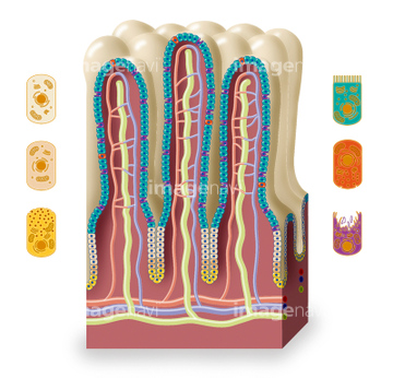
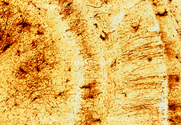
この検索結果には、Small intestine villi, section、Intestinal lining, SEM、Intestinal ulcer, light micrograph、Microvilli and a goblet cell、Intestinal microvilli, TEM、False-colour SEM of bronchial epitheliumなどが含まれています。
64251226
64049911
64206819
64251229
64196734

64225083
64049910
64116645
64055781
64060201
64060202
64234476
64234477
64234478
64234479

64225133
64234667
64234718
64056746
64147742
64021537
64055763
64152547
64209800
64213913
64213914
64213915
17202471
17202472
64224937
64060933
64075661
64021570
17201424
64073343
64021539
64021540
64021541
64021571
64225094
64021545
17201425
64088343
64196715
64090479
64090480

64225140
64056271
64073285
64021549
64021550
64021551
64161677
64161678
64161681
64161683
64161684
64072010
64072070
64072071
64021568
64021553
64116647
64150338
64049912
64224843
64057593
64057594
64057595
64057596
64057597
64057598
64073311
64132258
64132996
64132997
64021564
64021565
64021566
64021567
64234473
64234474
64150226
64150227
64234509
64234510

64088314
64090152
20536138
20536141
64021534
64021536
64066076
64071989
17212241
64061536
64247696
64021575
64264588
64264589
64040598
64040602
64077368
64021577
64021578
64021579
64021581
64207219
64207239
64207274
64224938
64197487
64197488
64021569
64199182
64021548
64021552
64209773
64209785
64209786
64211723
64211724
64211725
64002984
64002985
64060203
64060204
64190895
64021528
64050457
64071876
64071877
64073304
64088319
64088323
| 次ページ |
