HOME > ژتگ^ > ƒ‰ƒCƒtƒXƒ^ƒCƒ‹ > ‚¨ڈj‚¢ژ–پE’¢ژ– > ”DگPپEڈoژY
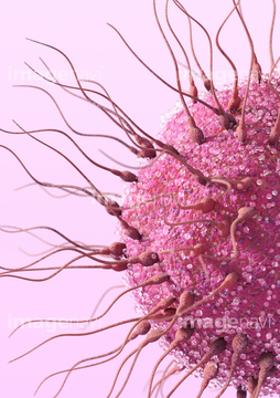
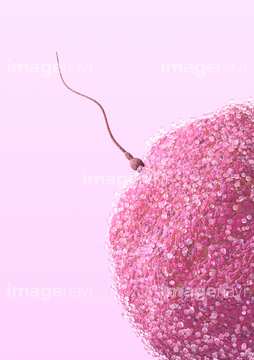
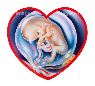
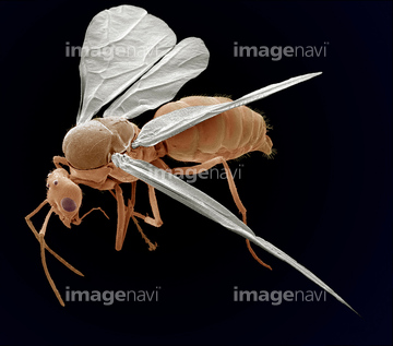
10,000Œڈ‚جژتگ^‘fچق‚ھŒںچُ‚³‚ê‚ـ‚µ‚½پB
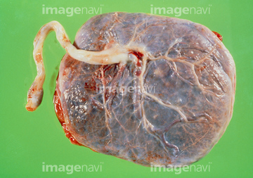
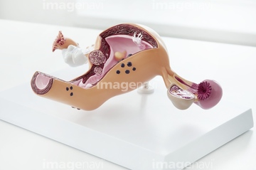
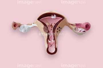
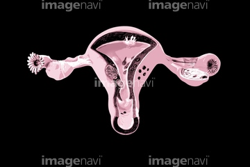
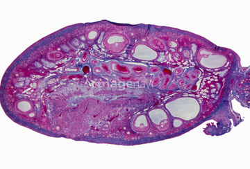
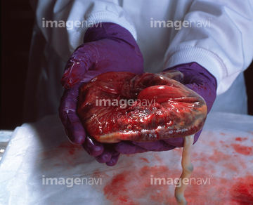
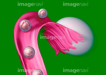
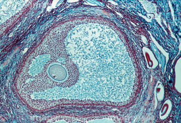
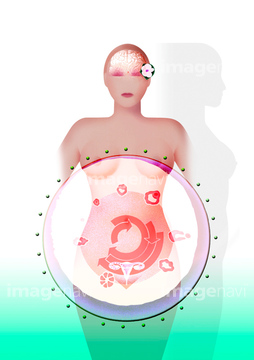
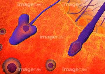
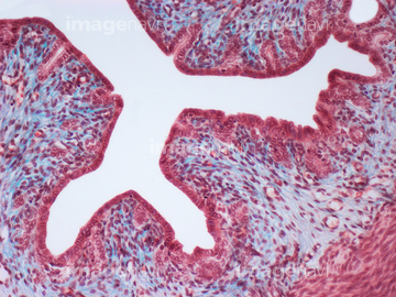
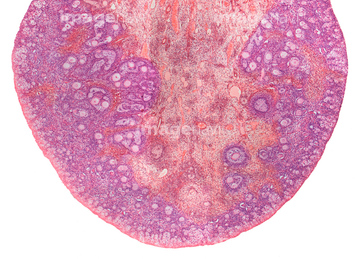
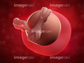
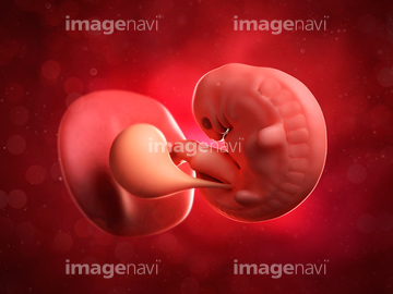
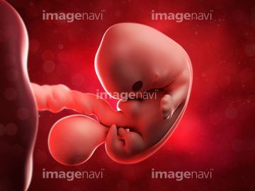
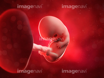
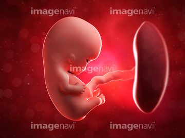
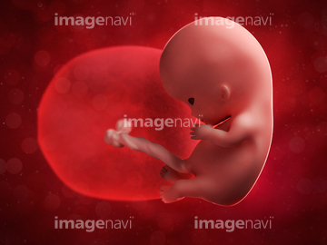
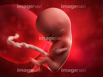
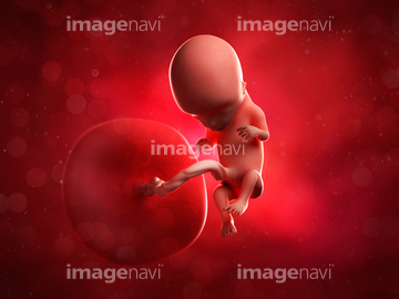
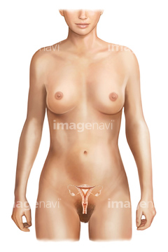
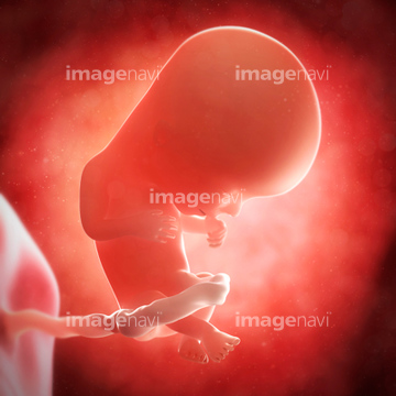
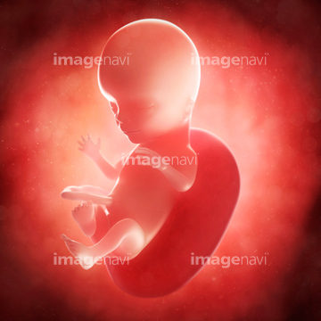
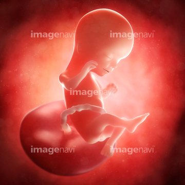
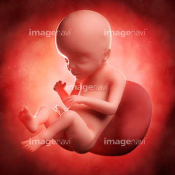
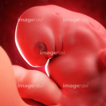
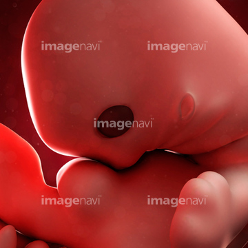
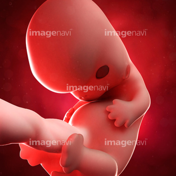
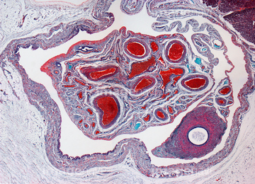
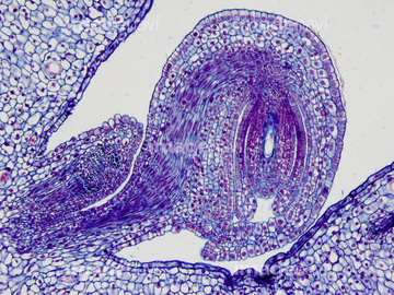
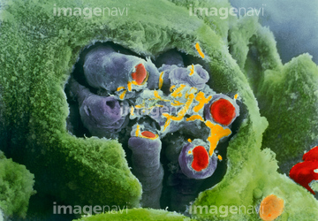
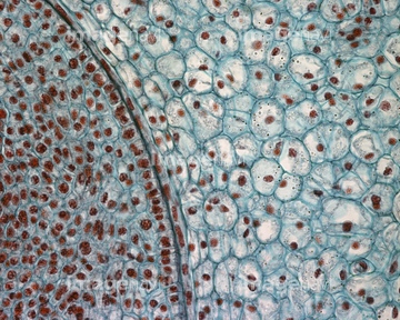
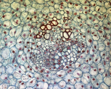
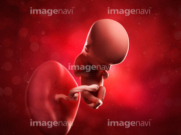
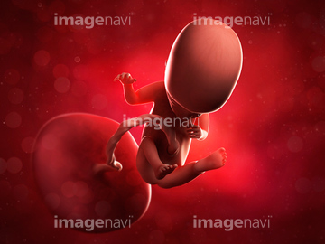
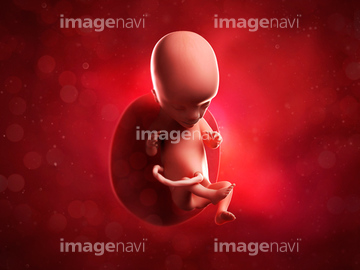
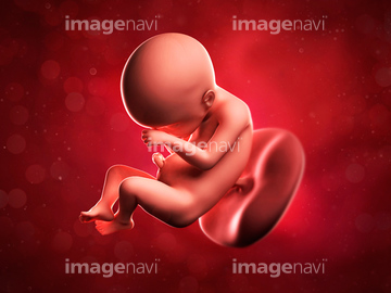
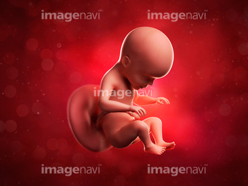
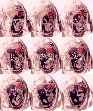
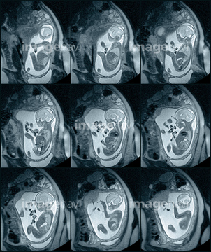
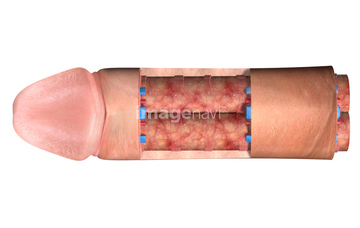
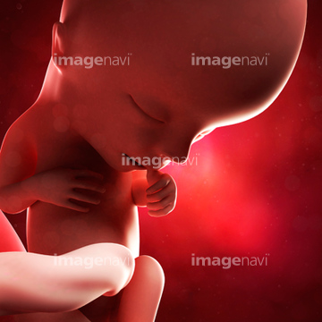
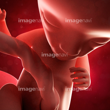
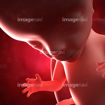
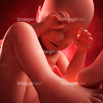
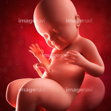
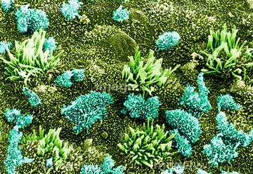
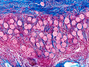
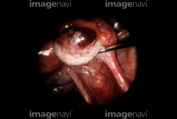
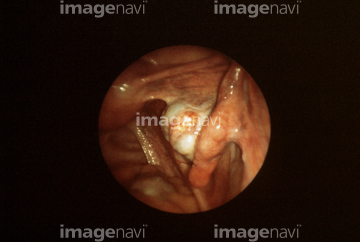
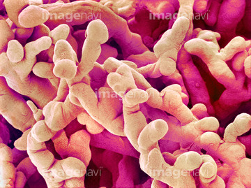
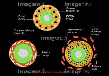
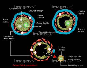
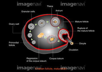
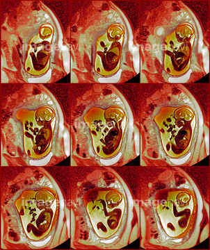
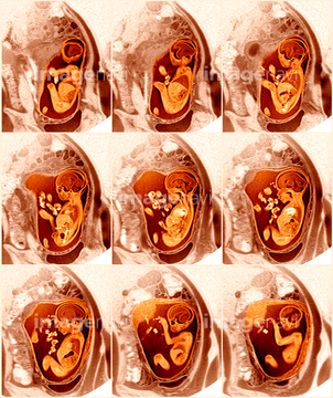
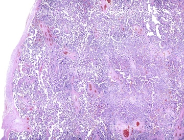
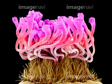
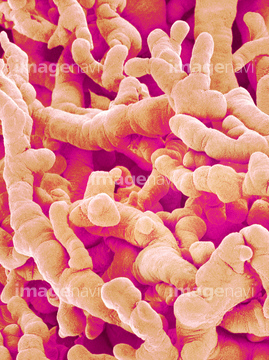
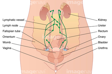
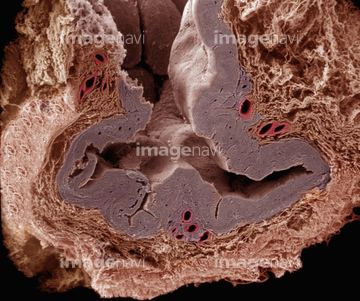
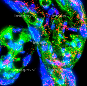
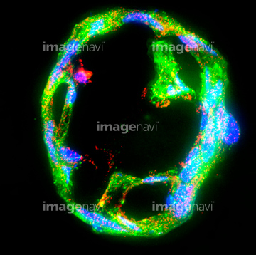
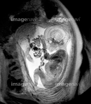
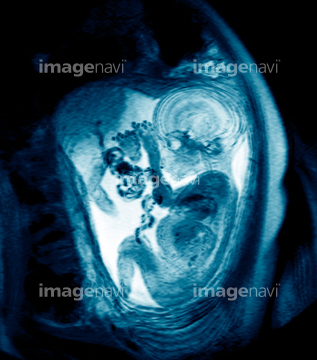
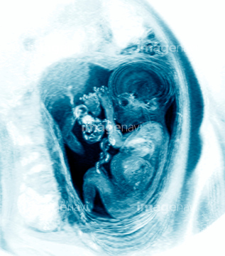
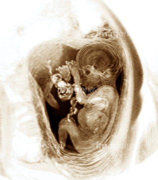
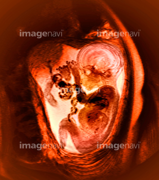
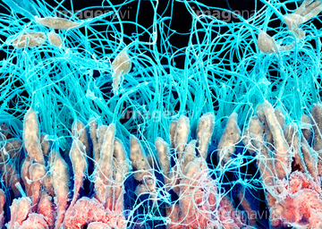
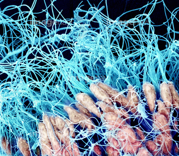
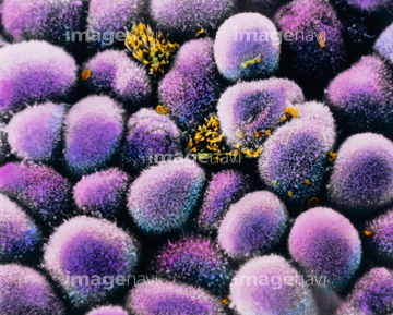
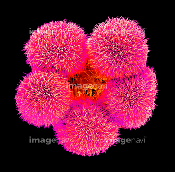
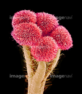
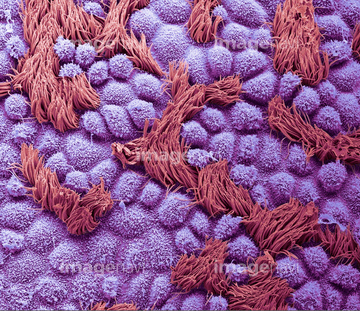
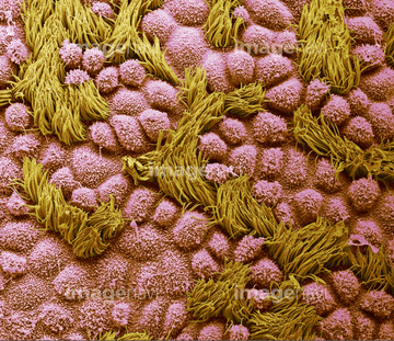
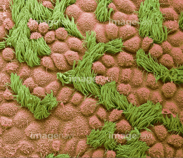
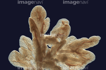
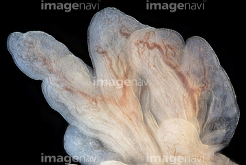
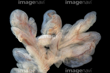
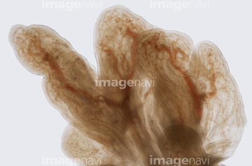
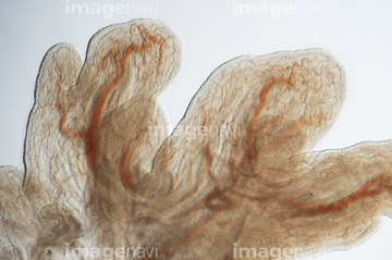
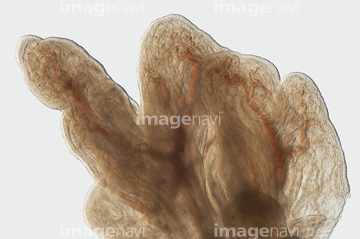
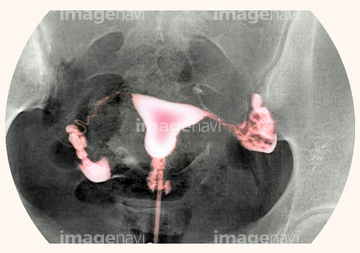
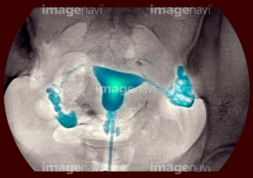
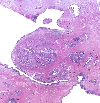
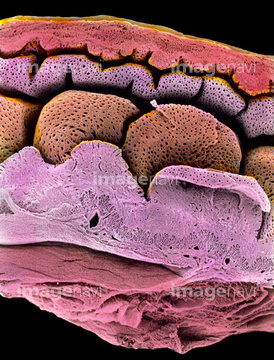
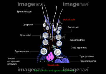
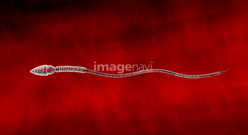
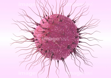
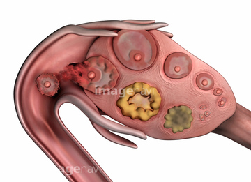
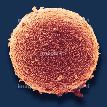
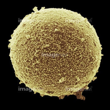
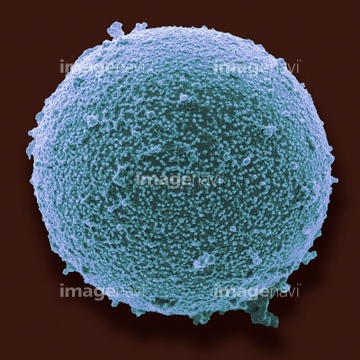
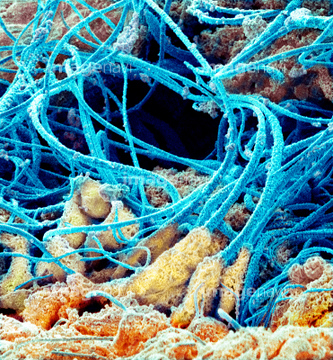
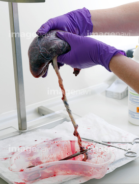
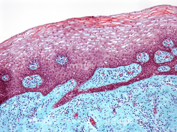
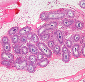
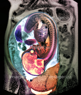
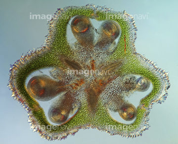
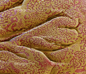
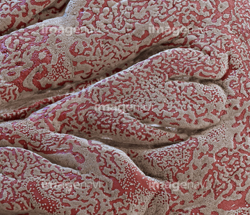
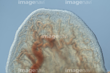
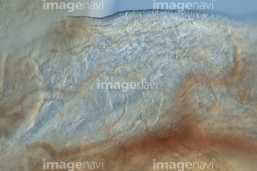
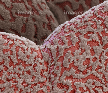
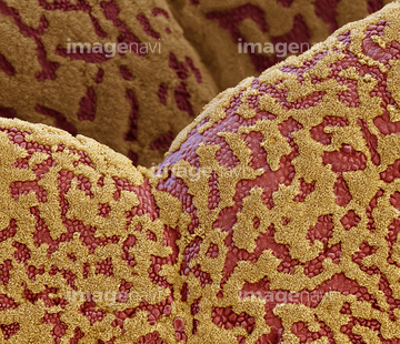
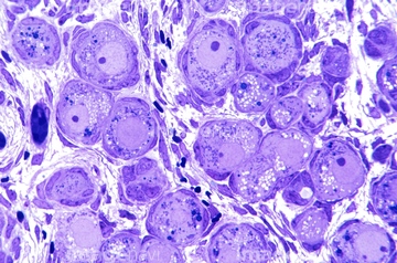
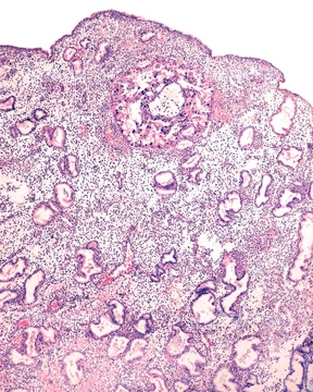
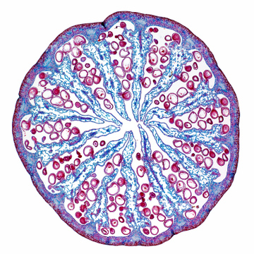
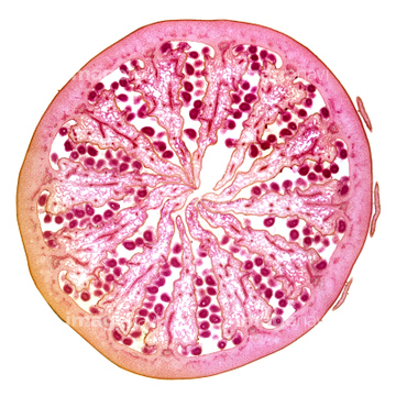
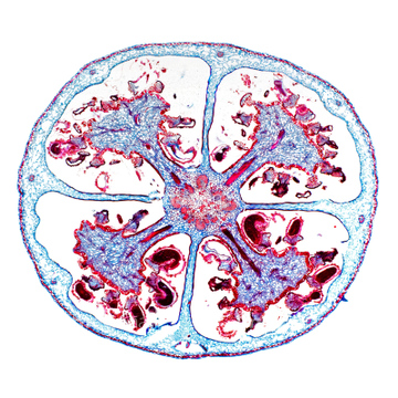
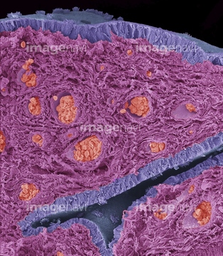
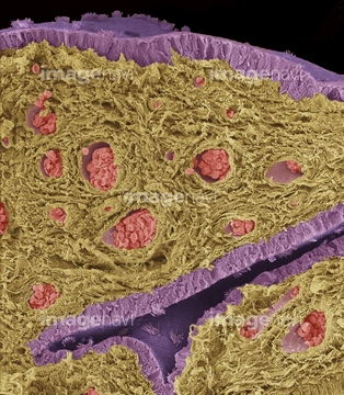
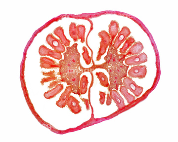
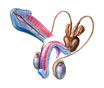
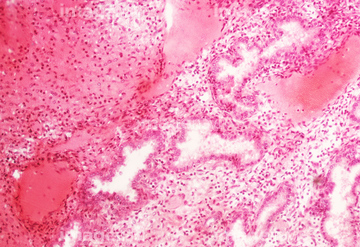
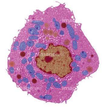
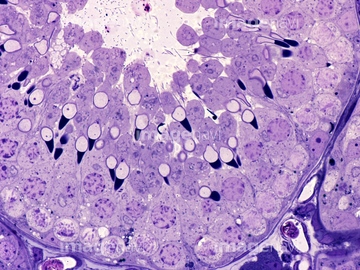
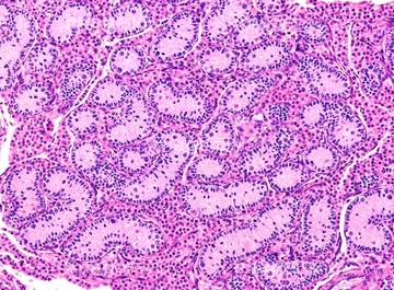
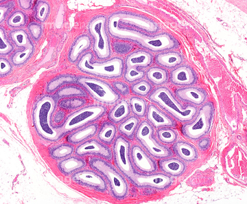
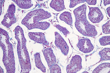
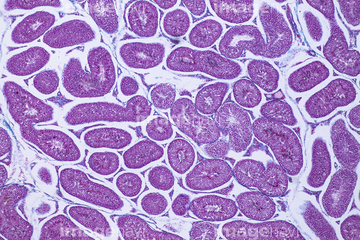
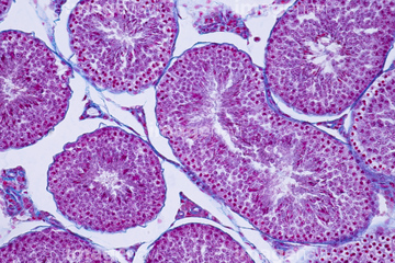
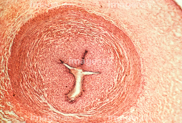
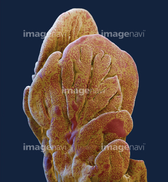
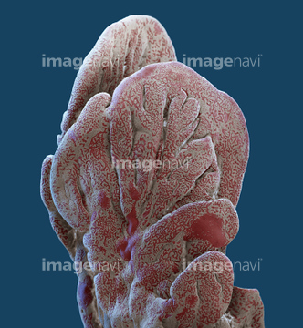
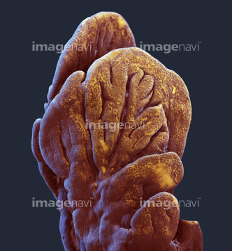
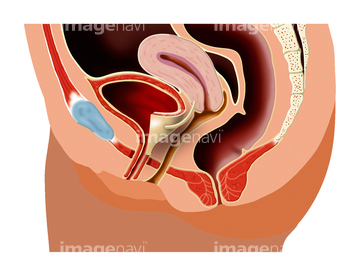
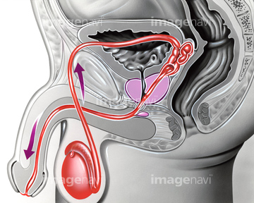
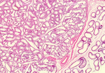
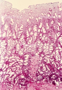
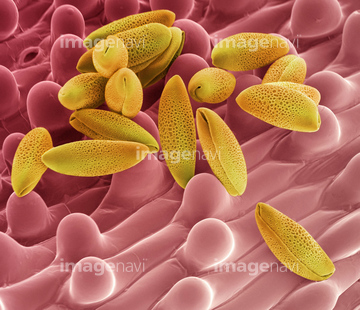
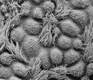
‚±‚جŒںچُŒ‹‰ت‚ة‚حپAFoetus at 26 weeks, artworkپAFoetus, MRI scansپAFalse-colour TEM of a human spermپAOvary, light micrographپAFlower's reproductive structures, artworkپAPenis arousal anatomy, artwork‚ب‚ا‚ھٹـ‚ـ‚ê‚ؤ‚¢‚ـ‚·پB
64068349
64021746
64067714
64067710
64021745
64051054
64093992
64088772
64058357
64049707
17212686
17212687
17212688
17212690
17212692
17212694
17212696
17212698
17212700
64070195
17211353
17211354
17211355

17211356
17211357
17211358
17211359
17211360
17211361
17212689
17212691
17212693
17212695
17212697
17212699
17212701
64093999
64263386
64021732
17212702
17212704
17212706
17212713
17212715
64021987
64021988
64060682
64098225
64059429
17212703
17212705
17212707
17212708
17212709
17212710
17212711
17212712
17212714
17212716
17212717
17212718
17212719
64060928
64021748
64021749
64021750
64021751
64021752
64021753
64021777
64049019
64049021
64049023
64021989
64021990

64063028
64067700
64076341
64021776
64050209
64225202
64008169
64008174
64019429
64019430
64019431
64021982
64021983
64021984
64021985
64021986
64076290
64077495
64060681
64076340
64077491
17290287
17290293
17290301
64021762
64021763
64048788
64048364
64048926
64071965
64111600
64111608
64111633
64076289
64019426
64093975
64093976
64098229
64136245
64136255

64249536
64249548
64085234
64044894
64199723
64199724
64199740
64199741
64199742
64199743
64049978
64048927
64048928
64067991
64040529
64040530
64040531
64040532
64051867
64116657
64019428
64084006
64084007
64084016
64192873
64192878
64084012
64060073
17201434
64103884
64106072
64098234
64076566
20530822
20531032
20531033
20531052
20531449
20531450
64092975
64199737
64199738
64199739
64050450
64067988
64040467
64152541
| ژںƒyپ[ƒW |
