HOME > 写真 > 人物 > 外国人 > 子供
10,000件の写真素材が検索されました。
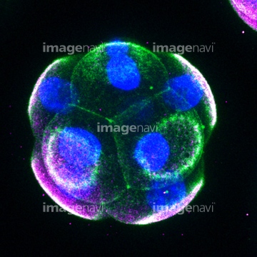
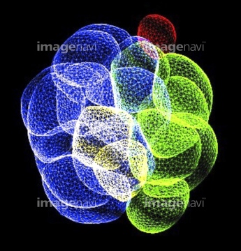
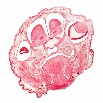
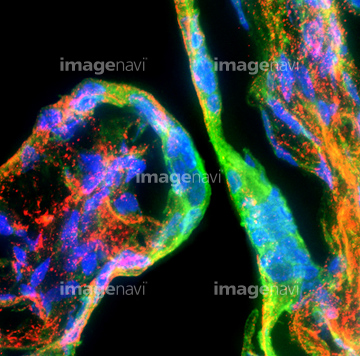
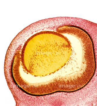
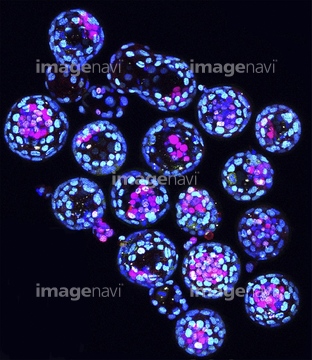
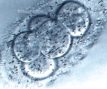
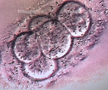
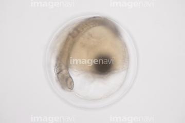
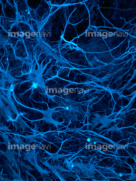
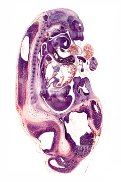
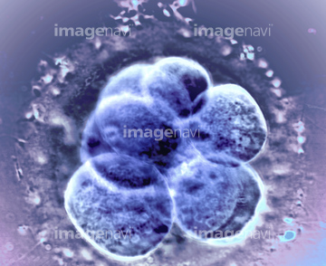
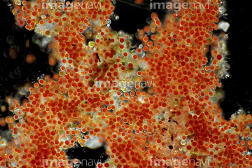
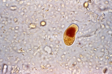
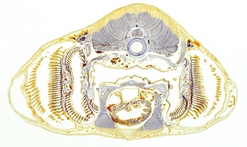
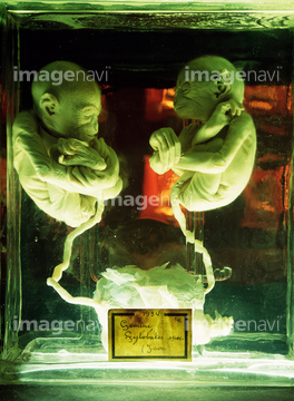
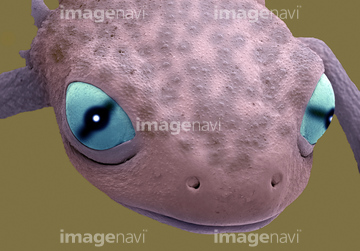
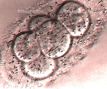
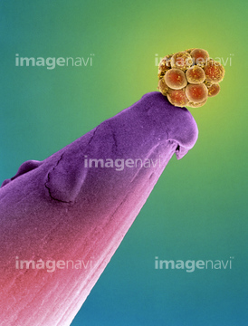
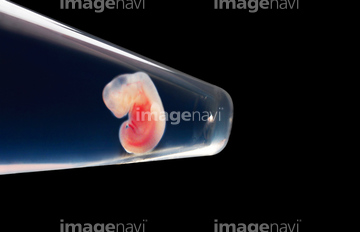
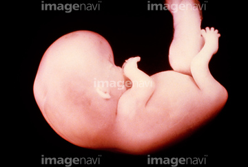
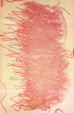
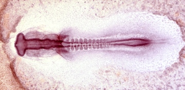
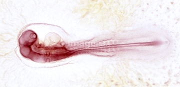
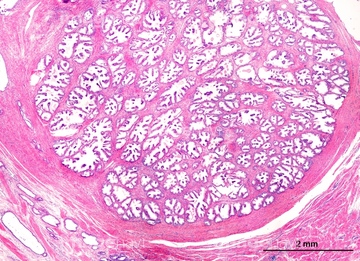
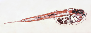
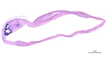
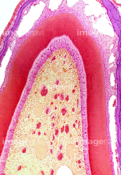
この検索結果には、Cat foetuses、Chicken embryo, light micrograph、Mouse embryo, light micrograph、Eight-cell mouse embryo, light micrograph、Sixteen-cell mouse embryo, confocal micrograph、Foetal rat head, light micrographなどが含まれています。
64068363
64071984
64071985
64071986
64063369
64063370
64056753
20568133
20568134
64225053
64119230
64190280
64022140
64049028
64058235
64073358
64010999
64084029
64084030
64022018
64022106
64225048
64244183
64244186
64244202
64190279
64022128
64022131
64224934
64010998
64022139
21553502
64225095
64118342
64049058
64049061
64224920
64073359
64022111
64022136
64022137
64063606
64063607
64067386
64022134
64022135
64085230
64085311
64085323
64044639
64022112
64022138
64245992
64245993
64086088
64152752
64044638
64067991
64083855
64083856
64022129
64022130
64124388

64108882
64108908
64041595
64060983
64060984
64060985
64047064
64071247
64071301
64152751
64022132
64022133
64085316
64152749
64060660
64124387
64089199
64261170
64261171
64261172
64261173
64107557
64034997
64033852
64245994
64152757
64072055
64089201
64163530
64119231
64058312
64071303
64152754
64129604
64072053
64213025
64213038
64213039
64156134
64156136
64257172
64209501
64209502
64063201
64180225
64043646
64012969
64116825
64116829
64149664
64149665
64129601
64129603
64129608
64129614
64129615
64129617
64044433
64244179
64244184
64244197
64012982
64168052
64168053
64168145
64168151
64168152
64188307
64188309
64188377
64188379
64158715
64158722
64172411
64190950
64190952
64190953
64076626
64022107
64063225
64063227
64063228
64063231
64008883
64008884
64088781
64124382
64152758
64092211
64009589
| 次ページ |
