HOME > 写真 > 科学・テクノロジー > 科学 > DNA・細胞
10,000件の写真素材が検索されました。
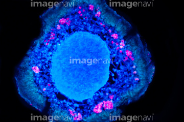
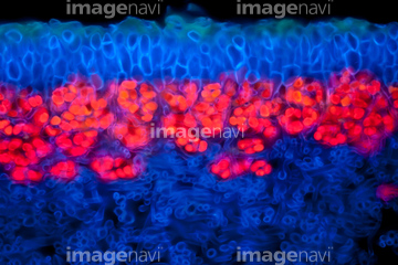
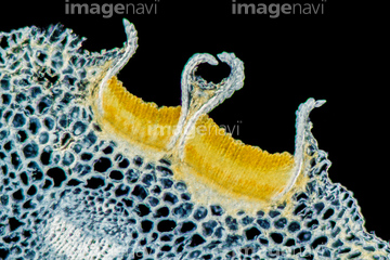
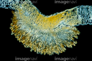
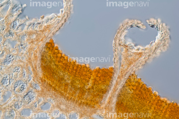
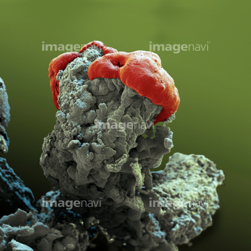
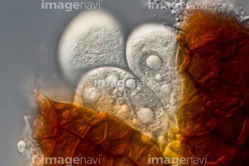
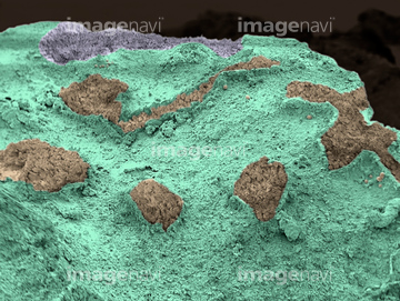
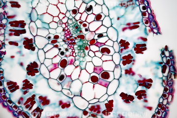
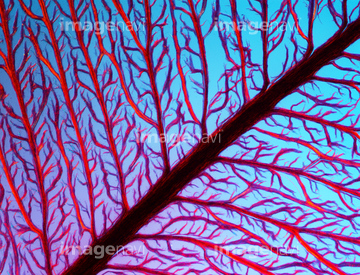
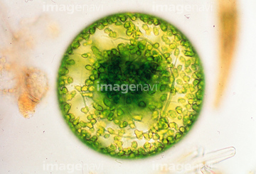
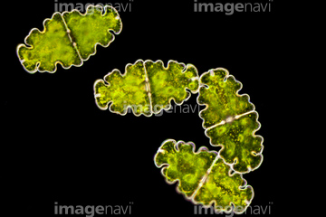
この検索結果には、LM of the red algae, Plumaria elegans、LM of the green alga, Eremosphaera、Euastrum sp. green alga, LM、Bambusina sp. green alga, LM、LM of a diatom alga, Navicula lyra、Verbascum sp. leaf, light micrographなどが含まれています。
64073057
64073058
64084210
64084226
64084217
64117362
64117381
64084181
64084184
64084185
64084205
64042114
64084224
64076226
64083991
64059733
64061093
64061094
64061095
64061114
64065495
17256246
17256253
64072711
64106488
64074679
64008091
64008094
64008097
64061537
64061538
64064828
64039726

64077400
64077414
64252154
64065498
64072698
64072708
64072710
64039733
64233701
64114534
64065508
64074677
64077363
64049249
64077415
64077416
64078129
64073056
64123505
64123513
64072695
64072700
64072703
64072705
64072709
64073054
64072683
64072685
64072687
64072688
64072690
64072692
64072694
64072696
64072697
64072699
64072701
64072702
64072707
64073055
64114533
64119749
64055756
64039756
64138219
64138223
64138243
64138248
64138256
64218412
64084173
64084200
64084201
64084219
64224934
64126830
64126833
64126840
64126841
64126845
64126847
64065486
64044433
64126828
64126834
20542515
17284286
64059685
64060767
64065504
64047088
64039732

64260076
64260100
64106203
64049248
64049250
64224931
64198303
64198317
64250405
64119728
64072691
64011725
20542485
20542486
20544117
64138232
64138247
64218394
64042918
64126839
64138221
64138236
64072706
64218384
64213976
64065490
64119708
64093466
64094094
64040202
17237098
17237099
17237100
17237101
17237102
17237103
17237104
17237105
17237106
17237107
| 次ページ |
