HOME > 写真 > 科学・テクノロジー > 科学 > DNA・細胞
10,000件の写真素材が検索されました。
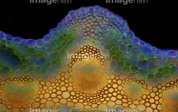
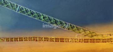
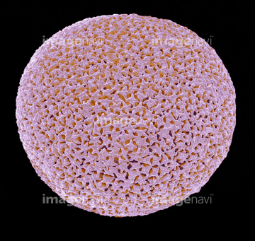
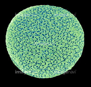
この検索結果には、Green hydra, light micrograph、Diatoms, light micrograph、Diatom, light micrograph、Plant trichome, fluorescent micrograph、Plant protoplast, fluorescent micrograph、Spirogyra algaeなどが含まれています。
64076417
64076409
64076443
64077457
64077458
64083993
64083997
64083998
64009116
64056946
64056947
64056948
64056949
64068393
64005640

64009115
64009118
64083992
64083994
64083995
64063704
64077449
64005641
64002854
64009025
64124825
64124833
64083996
64012409
64005660
64004043
64009047
64009048
64009049
64126577
64131575
20531024
64056791
64059696
64059697
64059698
64059699
64063689
64063700
64063722
64065499
64233503
64233504
64233690
64076429
64085021
64077447
64077448
64083622
64002864
64224846
64074896
64063702
64063717
64063720
64063723
64063724
64063725
64063726
64063727
64233688
64005662
64083734
64083735
64083737
64083738
64124814
64124821

64059691
64059692
64059693
64059695
64063721
64233505
64087493
64093489
64254812
64254890
64250239
64250242
64250244
64250245
64250575
64250581
64250582
64083619
64083742
64084481
64010748
64010749
64078793
64012870
64005401
64005403
64005404
64005405
64005417
64076304
64076309
64076312
64076313
64076314
64073436
64254897
64254898
64085398
64085401
64254793
17230542
17230543
64073633
64073635
64073637
64086076
64086077
64076088
64076318
64076319
64076410
64076411

64076415
64076418
64076421
64076422
64076470
64076471
64063610
64063611
64077315
64077316
64077317
64077318
64077319
64077320
64077321
64077358
64077367
64077383
64077389
64077390
| 次ページ |
