HOME > 写真 > 科学・テクノロジー > 科学 > DNA・細胞
10,000件の写真素材が検索されました。
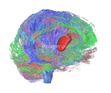
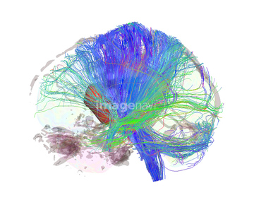
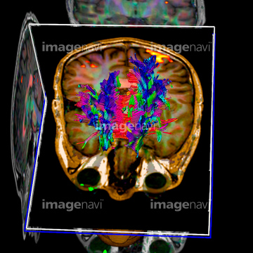
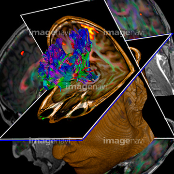
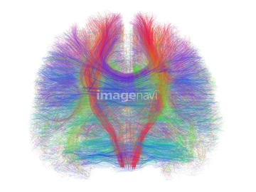
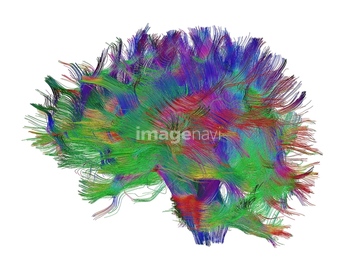
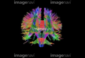
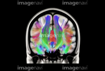
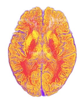
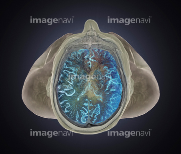
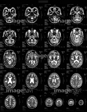
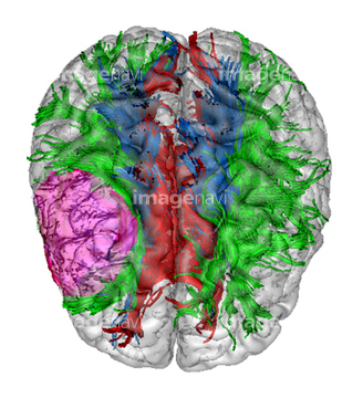
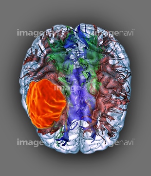
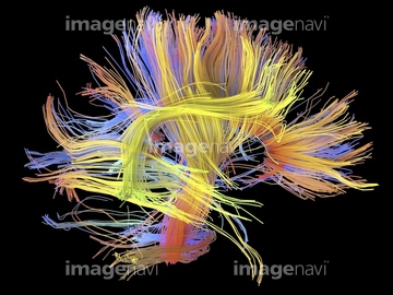
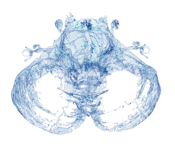
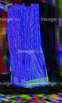
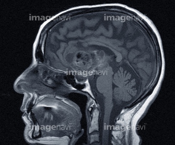
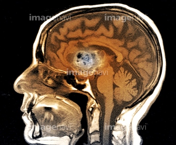
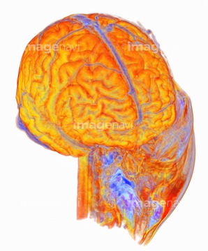
この検索結果には、Meningioma brain cancer, 3D MRI scan、Metastatic brain cancer, MRI scan、Human brain, 3D MRI scan、White matter fibres, brain mri scan、Brain tumour, DTI scan、Multiple sclerosis, MRI scanなどが含まれています。
64078080
64078081
64043117
64043118
64043119
64043120
64043121
64043109
64043110
64043111
64043112
64043125
64043128
64043129
64043130
64075025
64075026
64075027
64075028
64071941
64071942
64043139
17200425
64043086

64043087
64043088
64043089
64043090
64043097
64043098
64043099
64043100
64043101
64043102
64043103
64043104
64043105
64043106
64043107
64043108
64043131
64075564
64043091
64043092
64043093
64043094
64043095
64043096
64159968
64159969
64153281
64153282
64149562
64060094
64060095
64043132
64043133
64043134
64043135
64071938
64071940
64172494
64172495
64172496
64161158
64161159
64161160
64161161
64161162

64161163
64161164
64161165
64161171
64161172
64161174
64161176
64161177
64161178
64161179
64161181
64161182
64161183
64161187
64161194
64161200
64161201
64161202
64161204
64153283
64263862
64174843
64159954
64059701
64149559
64149565
64149566
64220586
64220585
64055604
64010056
64078939
64078940
64071925
64071926
64071927
64071447
64262287
64262289
64159971
64217040
64221200
64151226
64151227
64168002
64056411
64149563
64149564
64073310
64072632
64072640
64072641
64078941
64037601
64071706
64071707
64075285
| 次ページ |
