HOME > 写真 > 科学・テクノロジー > 科学 > DNA・細胞
10,000件の写真素材が検索されました。
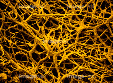
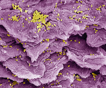
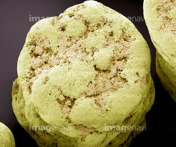
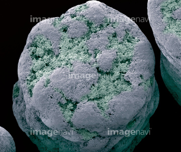
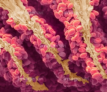
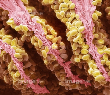
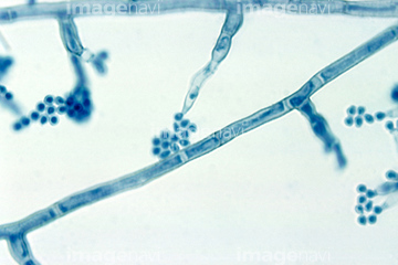
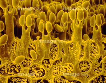
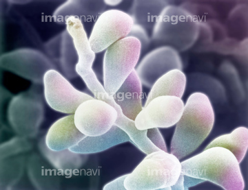
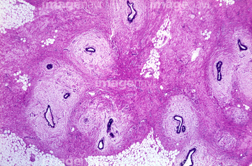
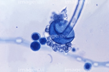
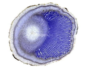
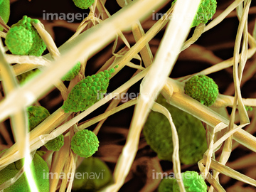
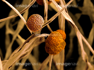
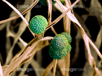
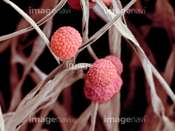
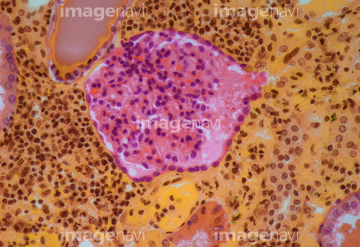
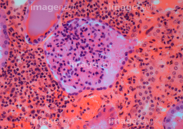
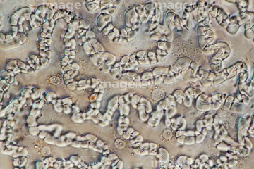
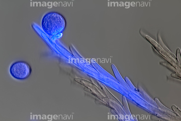
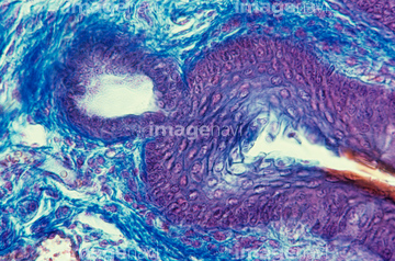
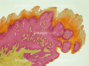
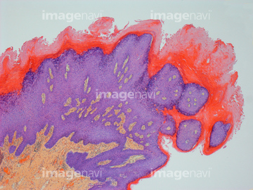
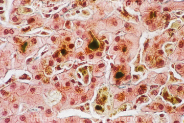
この検索結果には、Pholiota mushroom spores, SEM、Madurella mycetomatis fungus, light micrograph、Crumble cap of mushroom, Coprinus、Bread mould, SEM、Hepatocyte cells, light micrograph、Reticular cell, light micrographなどが含まれています。
64084217
64084226
64084184
64084185
64084210
64073058
64073057
64084181
64084205
64084224
64065495
64083991
17256246
17256253
64076226
64117362
64117381
64106488
64074679
64008091
64008094
64008097
64059733
64061093
64061095
64061114
64061537
64061538
64064828
64039726

64077400
64077414
64074677
64061094
64042114
64077415
64077416
64078129
64123505
64123513
64065486
64077363
20542485
20542486
20544117
64011725
64042918
64065490
17237098
17237099
17237100
17237101
17237102
17237103
17237104
17237105
17237106
17237107
17237108
17237109
17237110
17237111
17237112
17237113
17237114
17237115
17237116
17237117
64008092
64042119
64072768
64072783
64084170
64084173
64084176
64084186
64084195
64084200
64084201
64084203
64084219
64103836
64065111
64065112
64065113
64065114
64075671
64077379
64078818
64078895
64078896
64078916
64042924
64044433
64072711
64084178
64065498
64224934
64039733
64218871
64218872
64218873
64218874
64106489
64220255
64068369
64042122
64042123
64078130
64074777

64055756
64106203
64052277
64114581
64078566
64216546
64216547
64039663
64078568
64078569
64078573
20542508
20542515
20542529
17280701
17284286
64010272
64059685
64060767
64060929
64063012
64063013
64063014
64065504
64065508
64043727
64047088
64039651
64039652
64039653
64039655
64039656
64039732
64041415
64065874
64073323
64075077
64082277
64082278
64084495
64085266
64199170
64199171
| 次ページ |
