HOME > 写真 > イラスト・CG > 人物
10,000件の写真素材が検索されました。
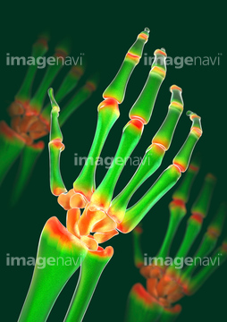
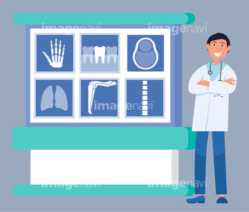
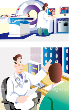
この検索結果には、Cervical spine, illustration、X-ray machine C-arm, illustration、Arthritic hand, X-ray artwork、Smiling doctor in uniform standing near x-ray boar…、MRIとCTスキャン、Human teeth, illustrationなどが含まれています。
64085431
64085430
64085432
64085445
64252466
64252467
64085596
64085405
64083954
64085447
64046492
51416744
64085408
64085604
64252461
64145408
64044528
64075436
64163428
64163429
64140070
64241936
64179295
64217191
64217192
64217193
64217194
64085605
64241922
51417425
64045675
16929678
64241958
64254265
64254266
64217556
64217595
64217596
64241923
64241934
51416745
64247222
64247223

64247224
64247225
64247226
64247227
64252407
64218378
64075965
31025048
64145365
64189640
64189641
64044518
20562428
20562436
20562437
51416920
41208273
64140071
17297970
17297971

17297972
17297973
17297974
17297975
17297976
17297977
17297978
17297979
20500771
20500786
20500859
20500914
20500926
20501000
20505678
20505679
20505863
20505864
20505975
20505976
20505988
20506551
20506645
20506650
20506739
20506741

20506743
20506744
20506745
20506749
20506778
20506780
20506781
20506945
20506946
20505323
20505324
20505325
20573670
20573671
20543351
20543373

20543376
20543419
20543420
20543430
20578775
20578778
20578783
20578784
64124647
64146809
64146810
64146811
64109752
64077221
64077222
64077223
64077224
64077225
64077226
64183425
64183443
64183444

64183445
64183447

64207574
64241930
64241938
64247538
64232261
64217787
64217821
64217822
64217823
64217824
64218047
64218048
64218049
64218050
64219113
64063136
64063137
64045975
64047189
64075437
64145381
41207202
20547136
41102478
41102479
41102481
64083974
64043376
20566734
20566729
20566730
20566731
20566732
20566733
64077229
20566888
31310591
64252460
64252462
15802957
17201824
17201825
| 次ページ |
