HOME > 写真 > 科学・テクノロジー > 科学 > 実験器具・装置
10,000件の写真素材が検索されました。
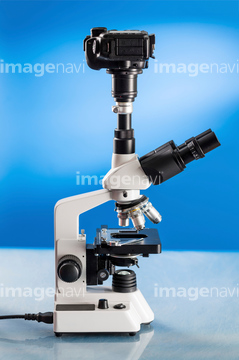
この検索結果には、光学顕微鏡、透過電子顕微鏡、産業・化学、顕微鏡とシャーレ、顕微鏡と葉、顕微鏡と実験道具などが含まれています。
64093270
10956009
10955973
17229691
17229692
17229693
64001091
99055136
10955994
30336133
10955937
99055137
19286443
14904446
99055033
64168930

17227175
17227230
17227231
17227292
17227280
64195232
17280771
17284298
17284414
17284415

17284416
17284417
20501172
14904444
99055031
99055032
99055034
30038529
64190286
10153992
10153993
10153994
17227212
17279129
17279130
10153996
10153997
20501171
64190315
64225608
99055134
20927653
64168931
99055135
17284282
17284283
17284284
17284285
64075985
64075986
64075987
64008571
16931438
16931439
10153991
17247640
17247241
64072724
64072736
99055140
10153995
10154003
64076066
64076067
64076068
64076069
64076070
64076071
64218334
64084173
64084219
64168932
64040806
64190316
64245053
17235614
17235615
99055133
64217290

30330145
17280701
64049905
64072735
64072774
64085241

64085243
64085244
64114552
64114571
64224937
64225203
64225212
64225213
64225224
64225247
20542485
20542486
20544117

64050011
64050018
17247212
64093272
64017885
64017886
64017887
64012330
64001232
64008653
64155162
64062612
64050014
64052278
64068369
16900161
16900162
64084761
17247236
17247239
17247243
17247244
64093250
64084484
64084487
64184913
64252788
64159839
64044433
64073057
64073058
64083975
64084200
64084201
64084210
64084226
64084495
| 次ページ |
