HOME > 写真 > 医療・福祉 > 医療 > 医薬品
10,000件の写真素材が検索されました。
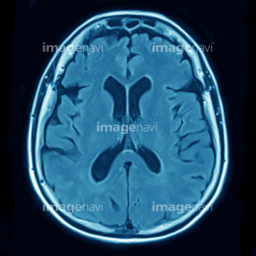
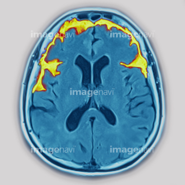
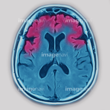
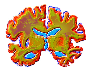
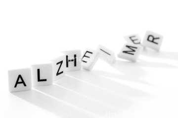
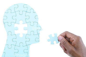
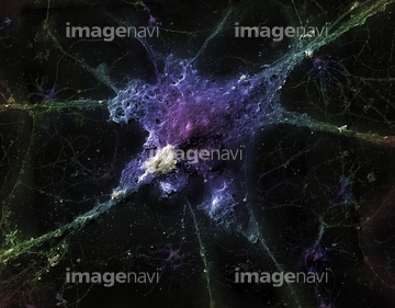
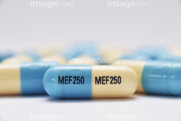
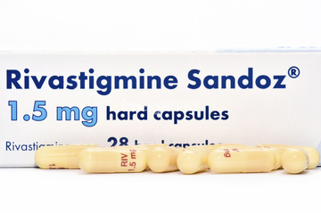
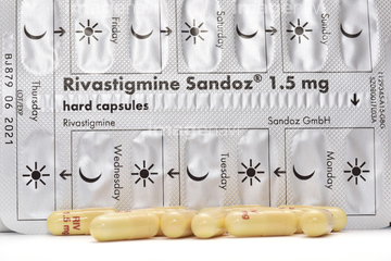
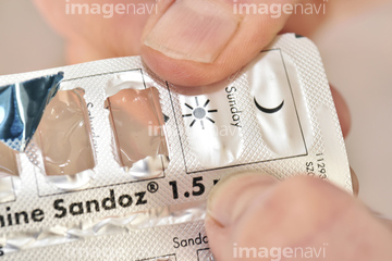
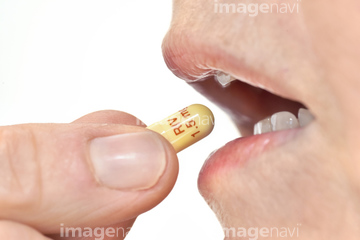
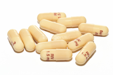
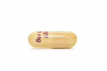
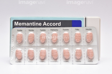
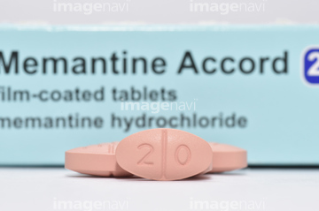
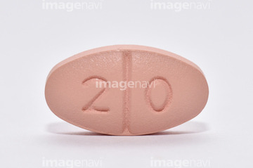
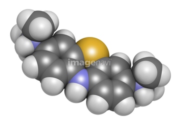
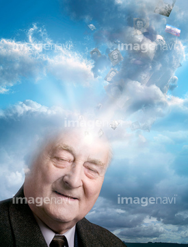
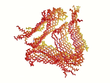
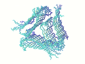
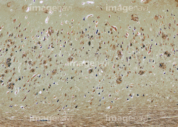
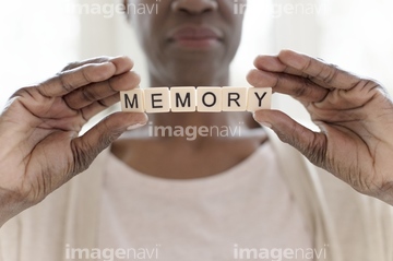
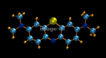
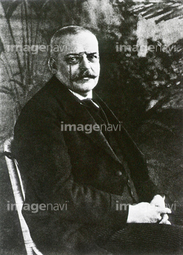
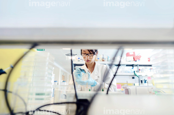
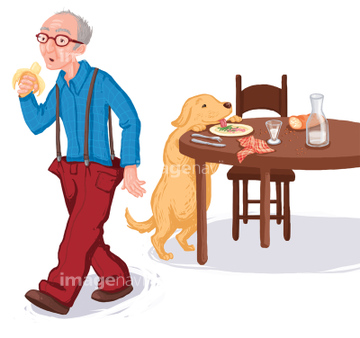
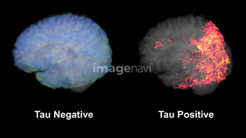
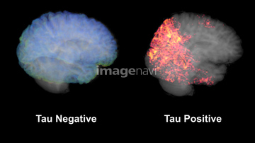
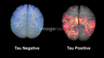
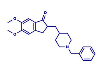
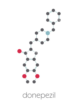
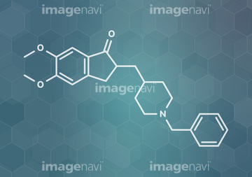
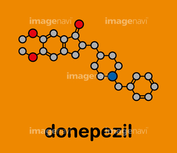
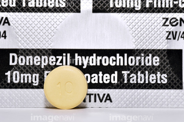
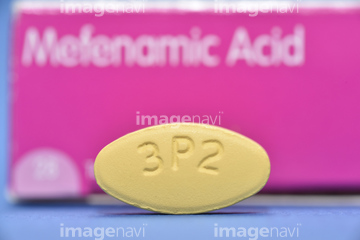
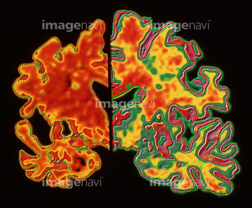
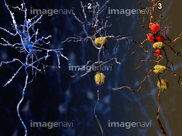
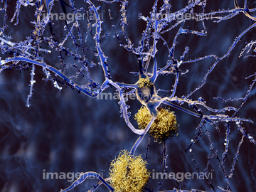
この検索結果には、Alzheimer's disease, conceptual image、Donepezil Alzheimer's drug、Mefenamic acid drug packaging、Mefenamic acid drug capsule、Mefenamic acid drug capsules、Rivastigmine dementia drugなどが含まれています。
64093957
64093958
64093956
64216528
64087036
53157256
53157257
53157258
53157259
64012918
64194355
64194356
64194357
64194358
64179314
64165630

64139123
53156548
53156549
53156556
53156557
53156558
53156559
53156560
53156561
53156562

53156563
64062611
64062617
64062624
64072631
64072644
53157029
20582428
64124609
64127299
64127301
64127302
64186898
64186899
64186900
64186901
64186902
64186903
64210309
64210310
64210311

64125399
64055054
64106797
64106799
64106801
64106802
64106805
64106806
64106807
64106808
64106809
64106810
17291210
17261256
20564681
64165637
64008418
64093963
53156979
64157116
64139124
64139125
64139126
64139114
64139120
64139121
20560090
64112235
64041266
64041267
64041268
64020504
20503310
17275974
17275975
17275976
17275977
17275979
17275980
17275981
17275982
17275984
20530785
20530786
20530787
20531339
20531340
20531341
20531342
20531343
20531344
20531405
20531406
20531407
64208814
64123113
64124610
64127300
64163408
64019879
64252142
64216199
64145861

64065734
64065735
64184039
17275891
17275893
17275906
17275907
17275910
17275911
17275978
17275983
64221065
17239188
17239189
17208414
17264976
17264977
64018481
64018484
17265136
17237929
64041269
64041270

17291209

17291211
17264982
17265184
64072416
17235297
17235298
17235299
64123111
64123112
17285015
17285016
20558229
20558231
64129528
64216159
| 次ページ |
