HOME > 写真 > イラスト・CG > 医療 > 健康
10,000件の写真素材が検索されました。
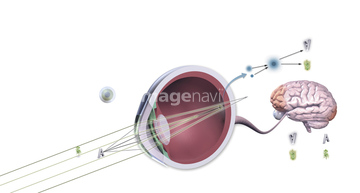
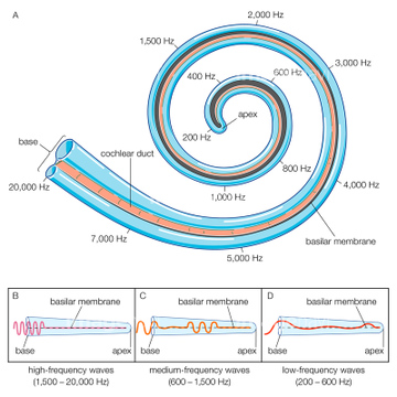
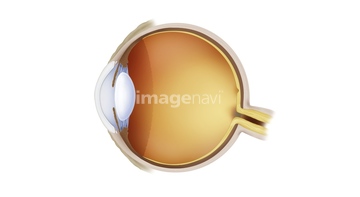
この検索結果には、耳の日、Labyrinthitis, illustration、Middle ear infection, illustration、Ear and cochlear anatomy, illustration、Evolution of the eye, artwork、Olfactory bulb in the brain, illustrationなどが含まれています。
64100259
64100260
64179285
64064269
64078913
64163513
64064270
64179286
17207857
64090014
64064268

64238645
64113536
64224220
64062956
64009872
16929677
64197089
17207859
64084946
64179255
64179254
64126085
64044882
17207858
64207398
64197085
64088297
64062170
64262694
40500445
64179252
64194841
64194842
64194843
64063128
64064093
64125928
64217402
64062344
64062169
64062171
64179356
64217530
64062342
64179253
64197211
64197268
64124648
64060081
64232082
64256877
64146593

64232097
64044818
64044881
20575528
20537829
20537830
20537831
20537832
64110038
64110041
64217871
64217872
64217873
64217874
64251628
64251629
64125929
64217780
40304882
40304883
64197208
64078912
64088457
64179284
64191005
14401679
64120689
64120690
64120783
64120784

64120785
64120786
64191819
64232091
64217790
64120762
64120763
64120764
64120771
64120772
64120773
64120774
64191817
64191837
64217176
64245279
20500549
20500769
20500778
20500784
20500810
20500856
20500865
20500881
20500944
20500974
20501018
20501040
64146578
64251630
64179295
64077217

20528337
64064125
64257515
64063146
64046494
64145365
64217781
64179298
64144466
64144467
64046458
20576156
20576157
20576162
20576163
20576164
20576165
20537733
20537734
20537735
20537736
20537737
20537839
20537840
20537841
20537842
20531966
20551682
17273436
20537796
20537797
20537805
64120691
64120692
64120701
64120702
64120746
64120747
64120759
64120760
64120761
64120767
64120770
64122557
64124647

64124649
64128640
64107616
64183412
| 次ページ |
