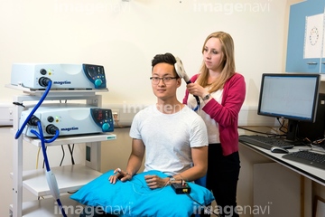
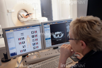
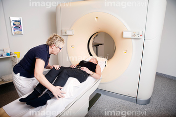
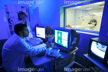
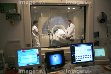
HOME > 写真 > 人物 > ビジネス > 学者・研究者
10,000件の写真素材が検索されました。
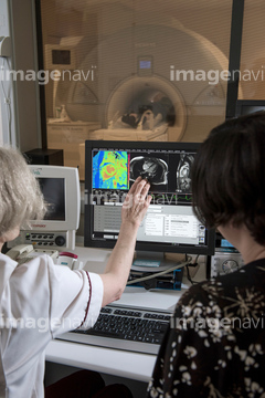
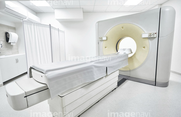
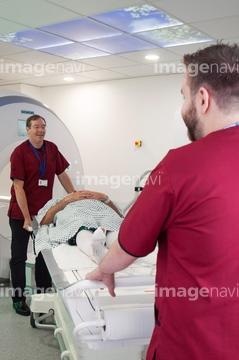
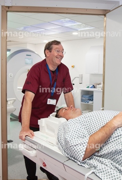
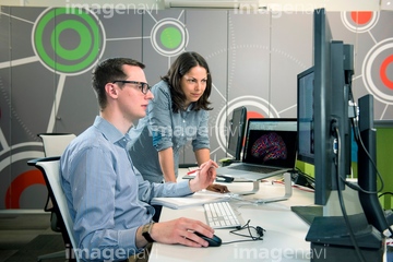
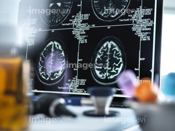
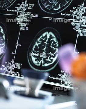
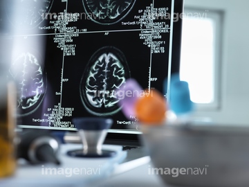
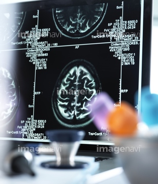
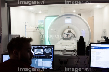
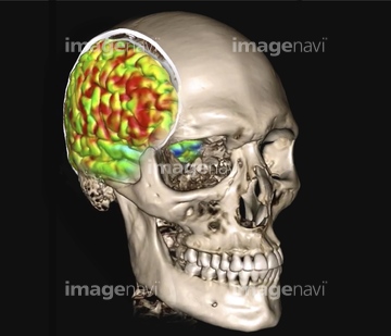
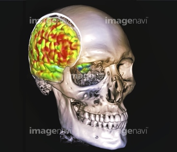
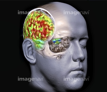
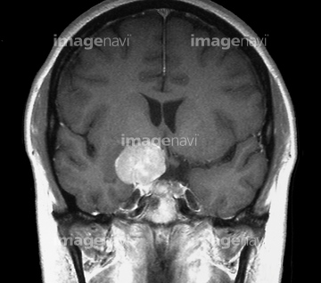
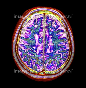
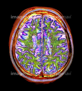
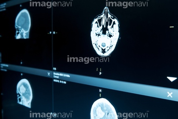
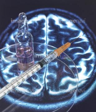
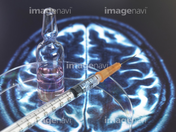
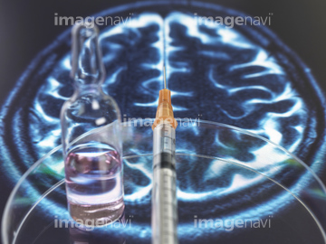
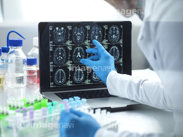
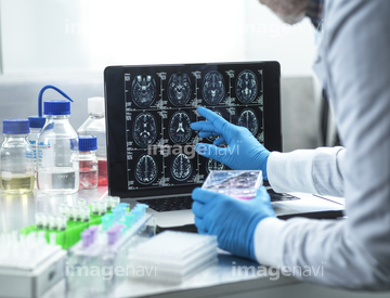
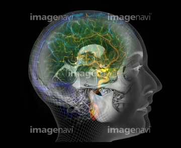
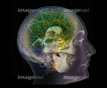
この検索結果には、Brain scans, MRI scans、Brain scan, MRI scan、MRI scan、Human brain's right hemisphere, fMRI scan、Human brain, fMRI scan、Human brain's left hemisphere, fMRI scanなどが含まれています。
64110131
64110129

64110151
64110152
64110153
64110154
64110149
64110150
64110134
64110135
64110146
64110147
64110148
64110133
64110136
64110138

20567839
20567840
20567841
20503377
64222838
20567674
64109657
64109658
64110137
17261836
17261837
17261838
17261839
64109014
64021466
64021467
17218191
17218192
17218197
17218200
17218204
17218205
17218208

17218210
17223829
17223837
17223840
64014403
64021474
64021475
64021477
64021478
64021479
64109656
64165282
64165283
20567603
17235936
17235937
17235938
17235939

64126440
64126441
64126442
64126443
64126454
64109000
64109001
64109015
64109016
64109017
64109019
64163208
64188151
64110139
21508369
64060094
64060095


20503373
64126330
64126331
17238389
64089178
64089180
64096033
64097113
64100636
64100638
64100639
64162334
64162335
64162336
64162337
64162338
64128197
64128204
64128205
40001439
40001440
40001441
40001442
17201739
17201768
17216126
17216129
17216136
17216148
17216193
17216195
17216196
17216206
17216208
17216211
64078939
64078940
64078941
64213180
64215014
64215028
64021464
17201406
17201407
17201776
64064532
64064533
64064534
64064535
64064536
64064537
64064538
64064539
64021476
64021481
20567601
64202000
17266349
17266353
17266357
17266360
20582752
20582753
20582754
64221444
64221448
64221449
| 次ページ |
