HOME > 写真 > 科学・テクノロジー > 科学 > DNA・細胞
10,000件の写真素材が検索されました。
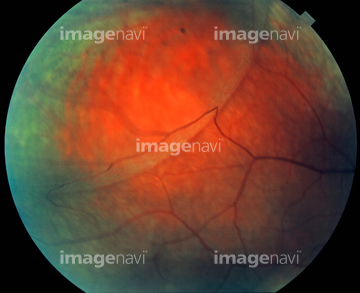
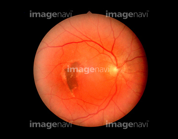
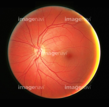
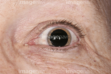
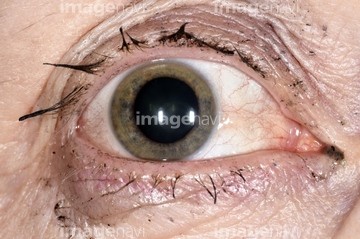
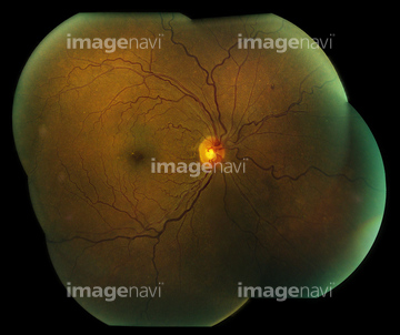
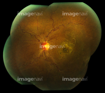
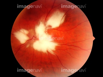
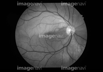
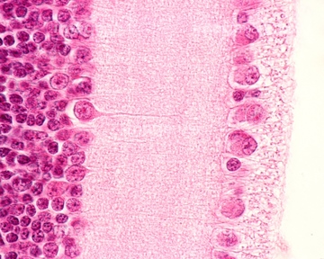
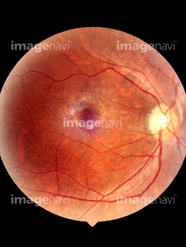
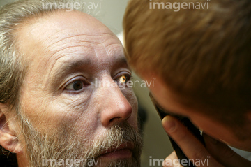
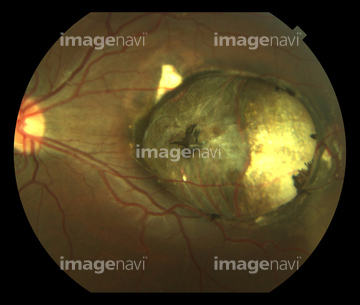
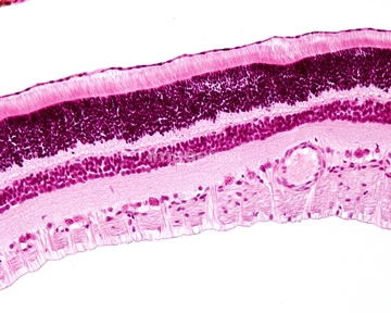
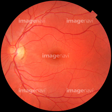
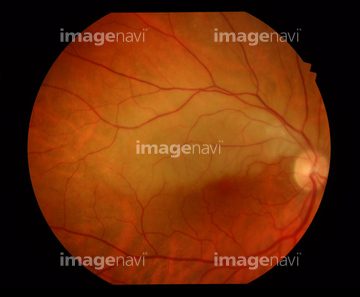
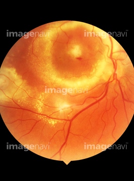
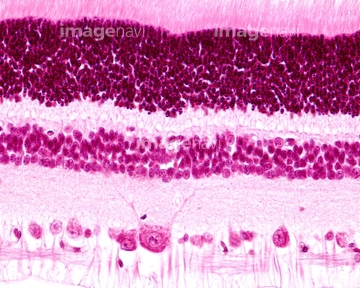
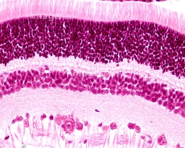
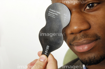
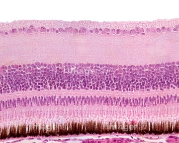
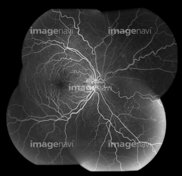
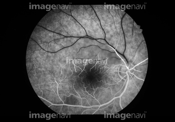
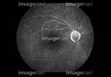
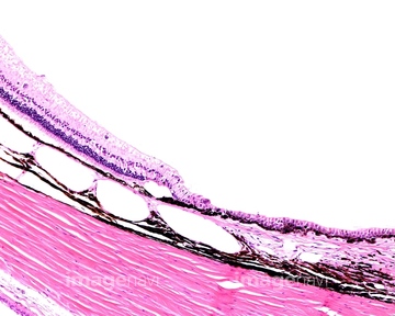
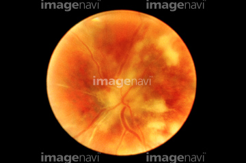
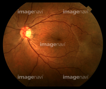
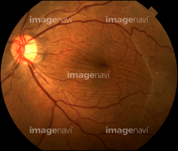
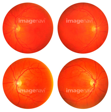
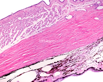
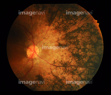
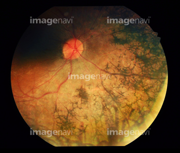
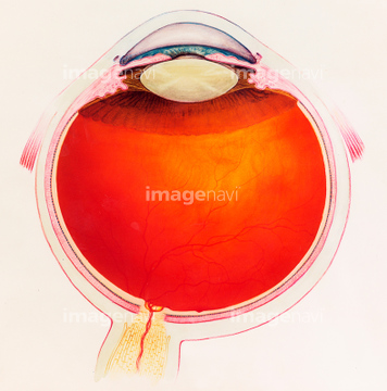
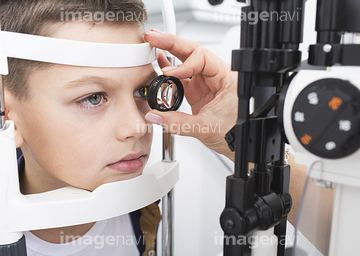
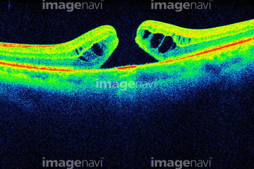
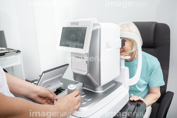
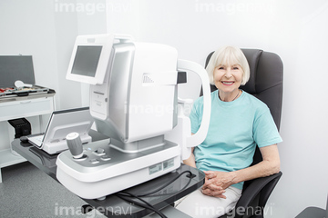
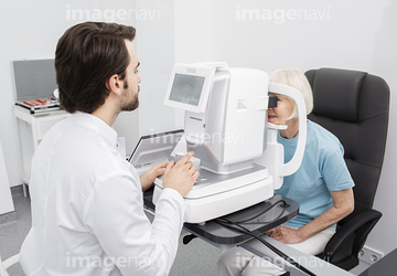
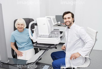
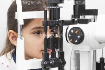
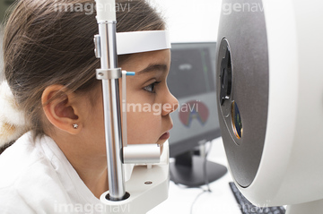
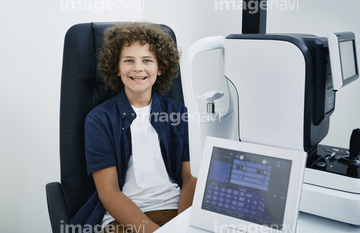
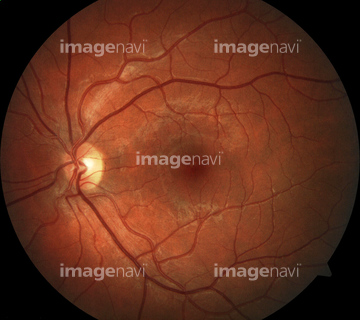
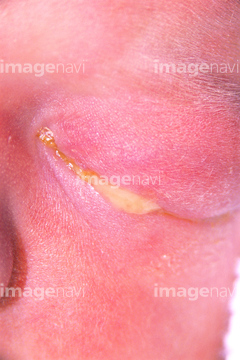
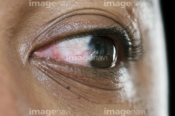
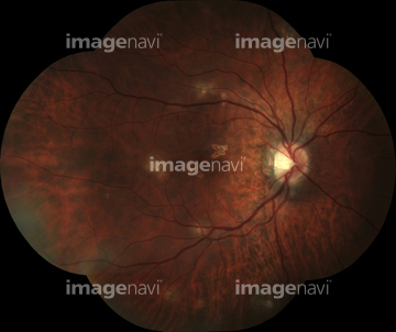
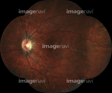
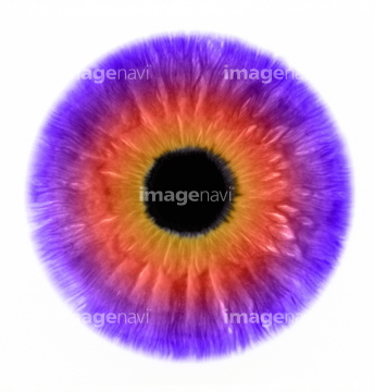
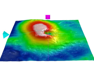
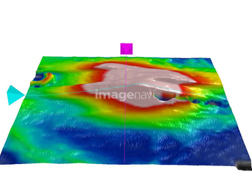
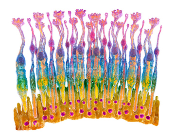
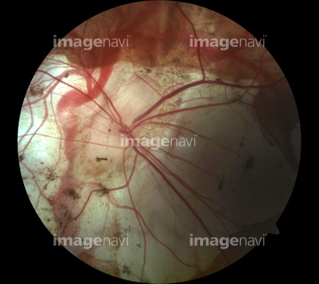
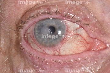
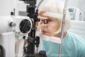
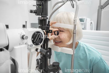
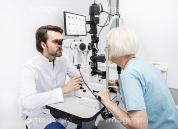
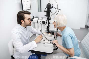
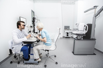
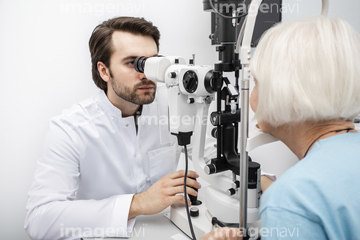
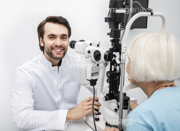
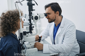
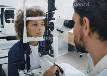
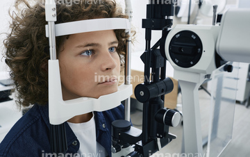
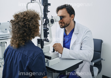
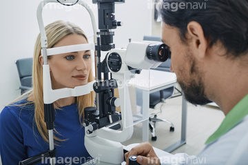
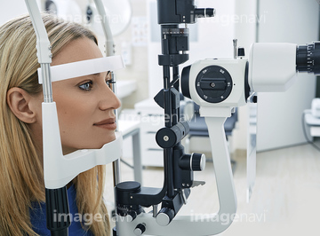
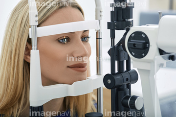
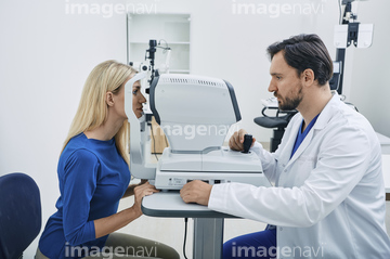
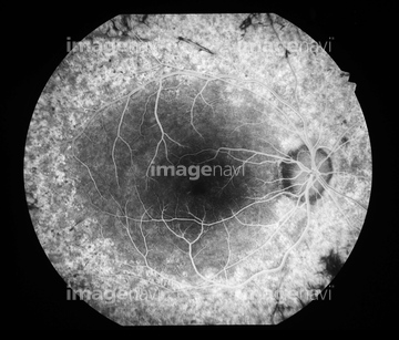
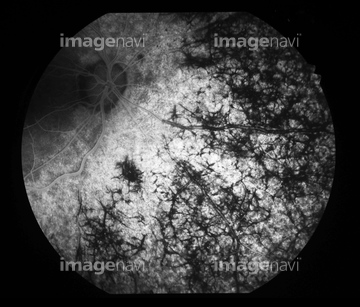
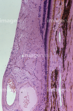
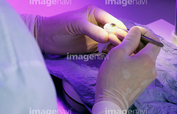
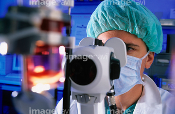
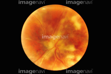
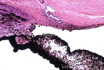
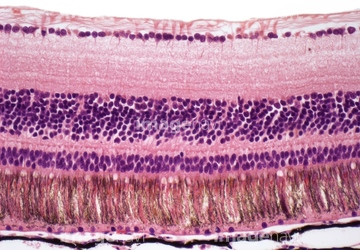
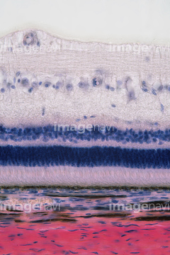
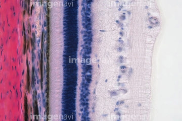
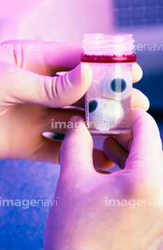
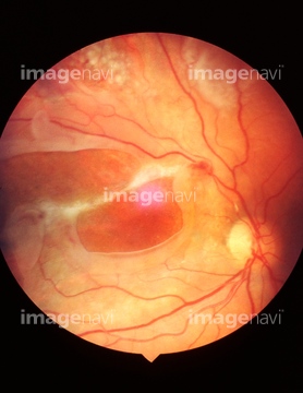
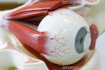
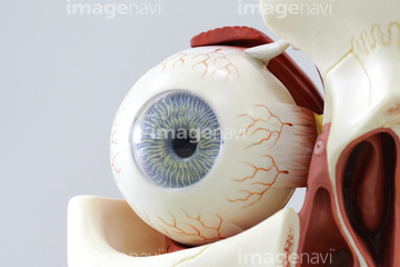
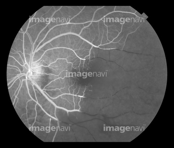
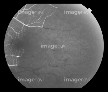
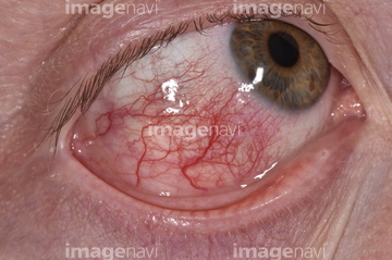
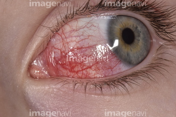
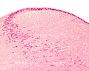
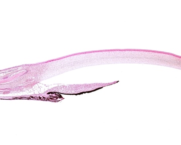
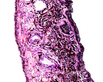
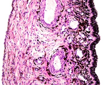
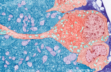
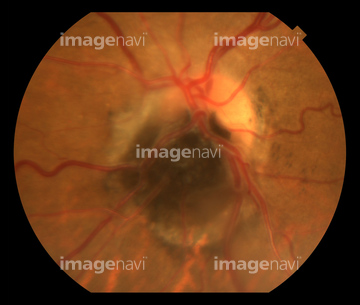
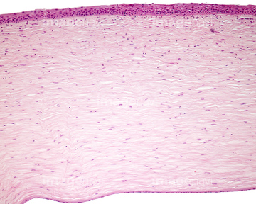
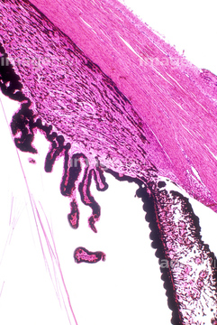
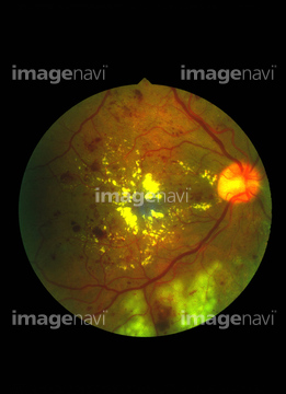
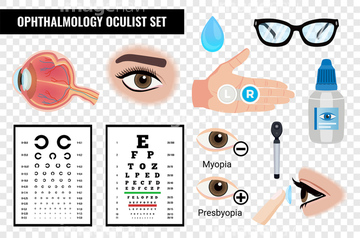
この検索結果には、Human eye for cornea harvesting、Eye examination、Retinal blood vessels, OCT angiography scan、Diagrammatic cross section of the human eye、Macular hole, OCT scan、Eye testなどが含まれています。
64114190
21511472
64168146
64190956
64247753
64247754
64109012
64019226
64247843
64198974
64109011
64198977
64198978
64243387
64216438
64216441
64216442
64260274
64190721
64190723
64190724
64190726
64038773
30324303
64216424
64248120
64248122
64248123
64248124
64038848
64248020
64248021
64247729
64040291

20549281
20560219
64247845
20549228
20549229
20549244
20549245

20560016
20560083
20560181
20567689
64248022
64040287
64040288
64188310
64153741
64153742
64167382
64167383
64163511
17225435
64102939
64102940
64188305
64188306
20549230
20549231
20549232
20549233
20549234
20549235
20549236
20549237
20549238

20549239

20549240

20549243
20560032
20560034

20560078

20560163
20560179

20560180
20560195
20560196
20560197
20560014

20562745

20562746
20566184
20566185
20566186
20566187
20566188
20566189
20566190
20566191
20566192
20566193
20566194
20567675
20567676
20567677
20567678
20567679
20567686
20567687
20567688
20567693
20567694
20567695
20567696

64019233
64248125
64248126
64040289
64040293
20532901
20567690
64235071
64235073
64235074
64235075
64235076
64040290

20532612
20532613

64102380
64247724
64247725
64093922
64225221
64177839
52202138
20531001
20531002
20531003
20531004
20531005
20531006
64188311
64188351
64194306
64190713
64263134
64188312
64260261
64224839
64151616
64216423
64216439
64216440
64056270

64040292
64247977
64260240
64122894

64247739
64248257
64248260
20532614
64115853
| 次ページ |
