HOME > 写真 > 科学・テクノロジー > 科学
10,000件の写真素材が検索されました。
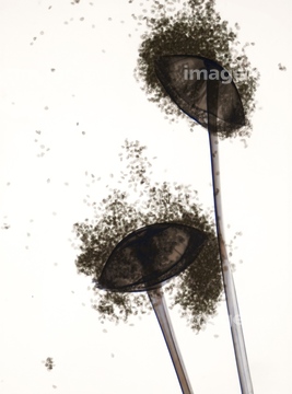
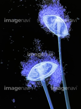
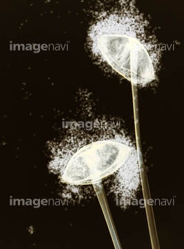
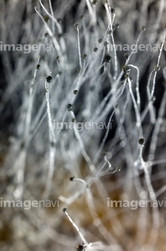
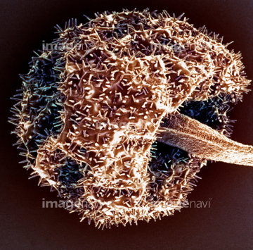
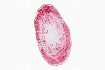
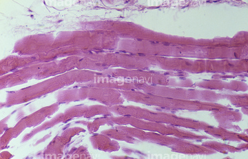
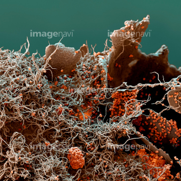
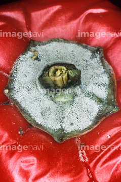
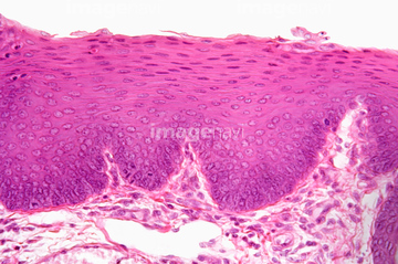
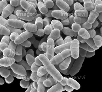
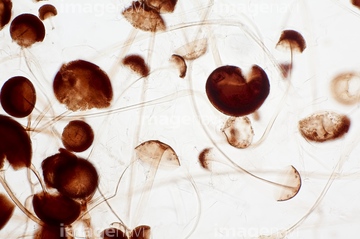
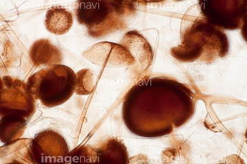
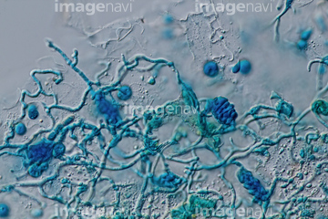
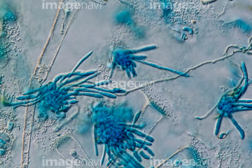
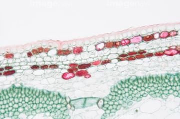
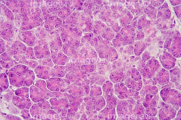
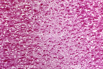
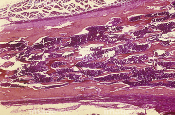
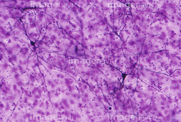
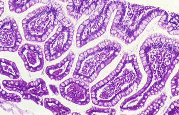
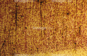
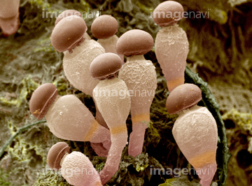
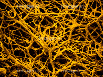
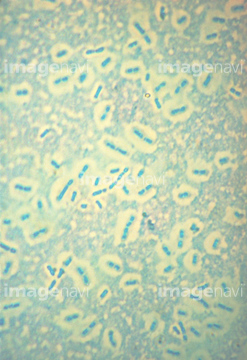
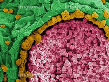
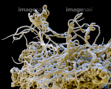
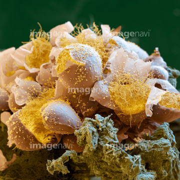
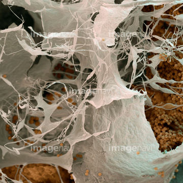
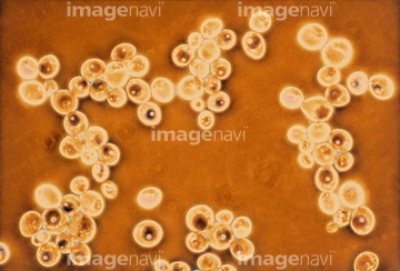
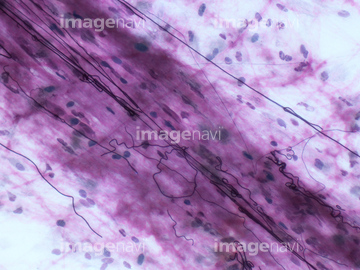
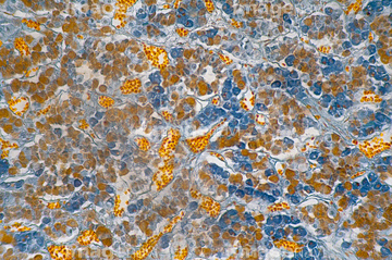
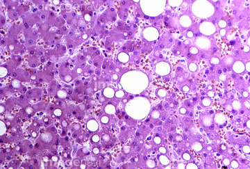
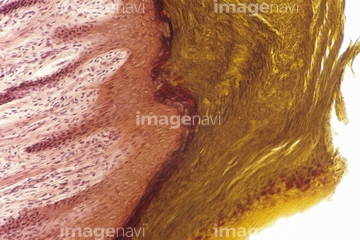
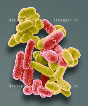
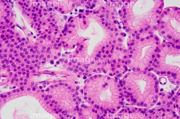
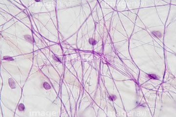
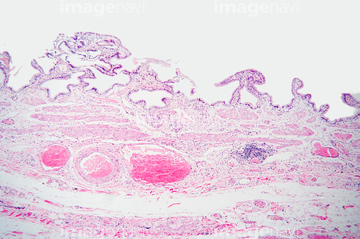
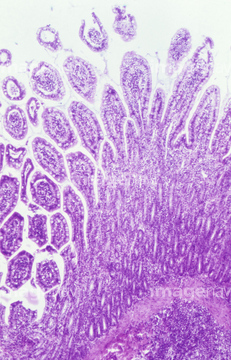
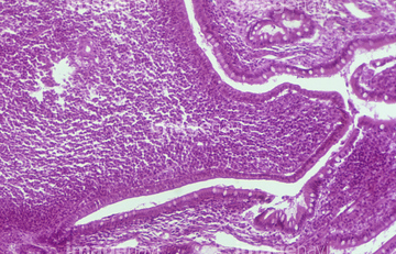
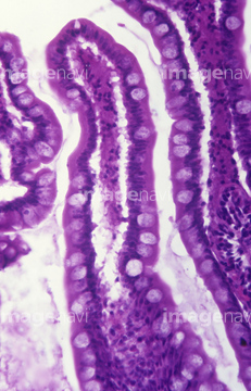
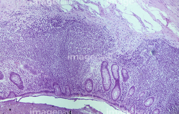
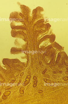
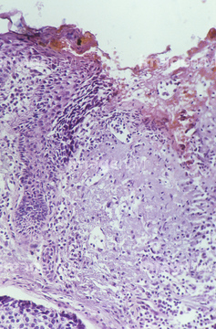
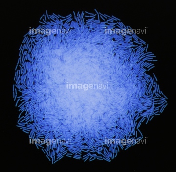
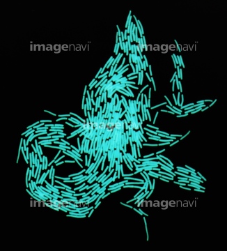
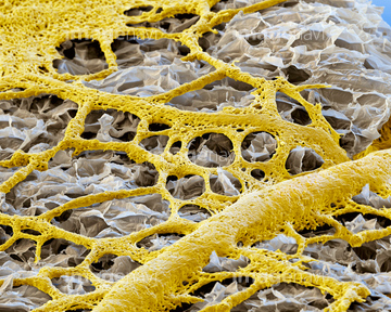
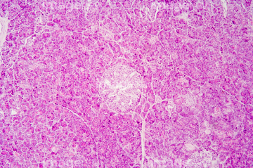
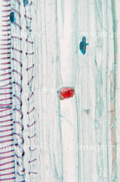
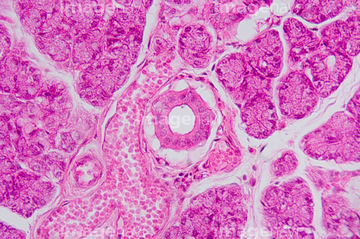
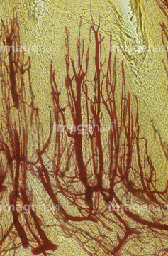
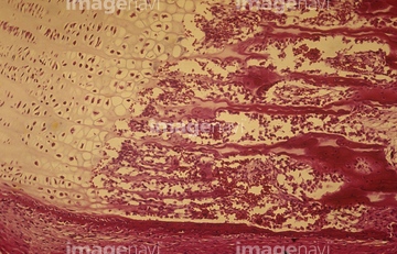
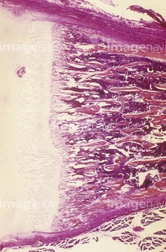
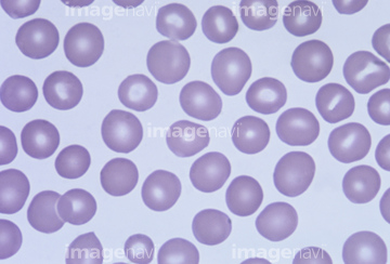
この検索結果には、アオカビ、Section of pancreas acinar cells. LM、Glycogen in liver tissue LM X160、Developing human long bone and marrow. LM、Human brain neurons with glia cells. LM、Villi of the human small intestine. LMなどが含まれています。
64114581
64224853
64225084
64225097
64003365
64106203
64003432
64077363
64003466
64074677
64003465
64114579
64003514
64003513
64003439
64003442
64003447
64003452
64003454
64014381
64218871
64218872
64218873
64218874
64101267
64003413
64003440

64003443
64003451

70014586
64003574
64114577
64003660
64003667
64003666
64114561
64152588
64114534
64114540
64224928
64003368
64074679

64225212
64003444
64224880
64059733
64003448
64003449
64003450
64065495
64088782
64206590
64115571
64225224
64078129
64198303
64198317
64114580
64115581
64224907
64224922
17246260
17246264
17246265
64154995

64003661
64003668
64250426
64114541
64114570
64114571
64114575
64224932

64224937
64224938
64224942
64225204
64225221
64225231
17256212
17256219
17256250
17256259
64114521
64224904
64003412

64003445
64003446

64225213
64225232
64225233
64225247
64225293
64138232
64138247
64138256
64224929
64254737
64254772
64250416
64217577
64114566
64224840
64224862
64225156
64225175
64123516

64123518
64123524
64029976
64216546
64216547

64224841
64224844
64084210
64084217
64084226
64224839
64073058
64220255
64003354
64003355
64003363
64003367
64003477
64003478
64011725
64178273
64178280
64178281
64003371
| 次ページ |
